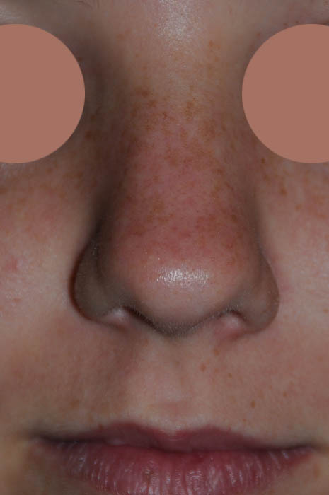
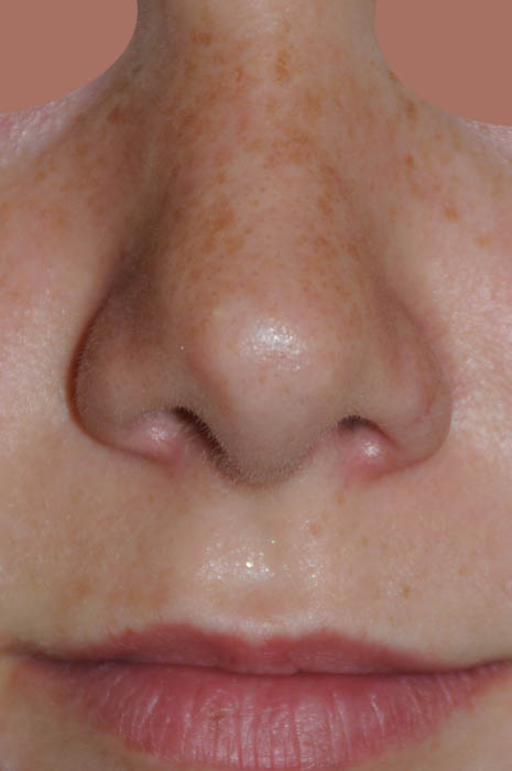
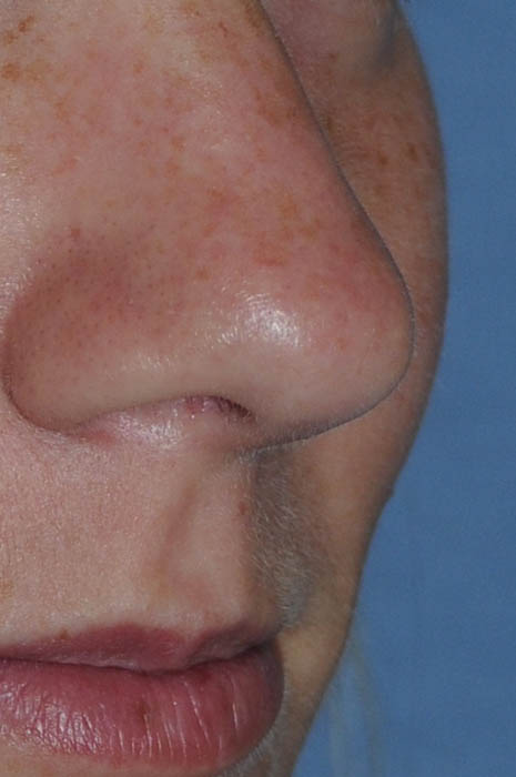
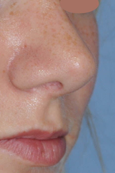
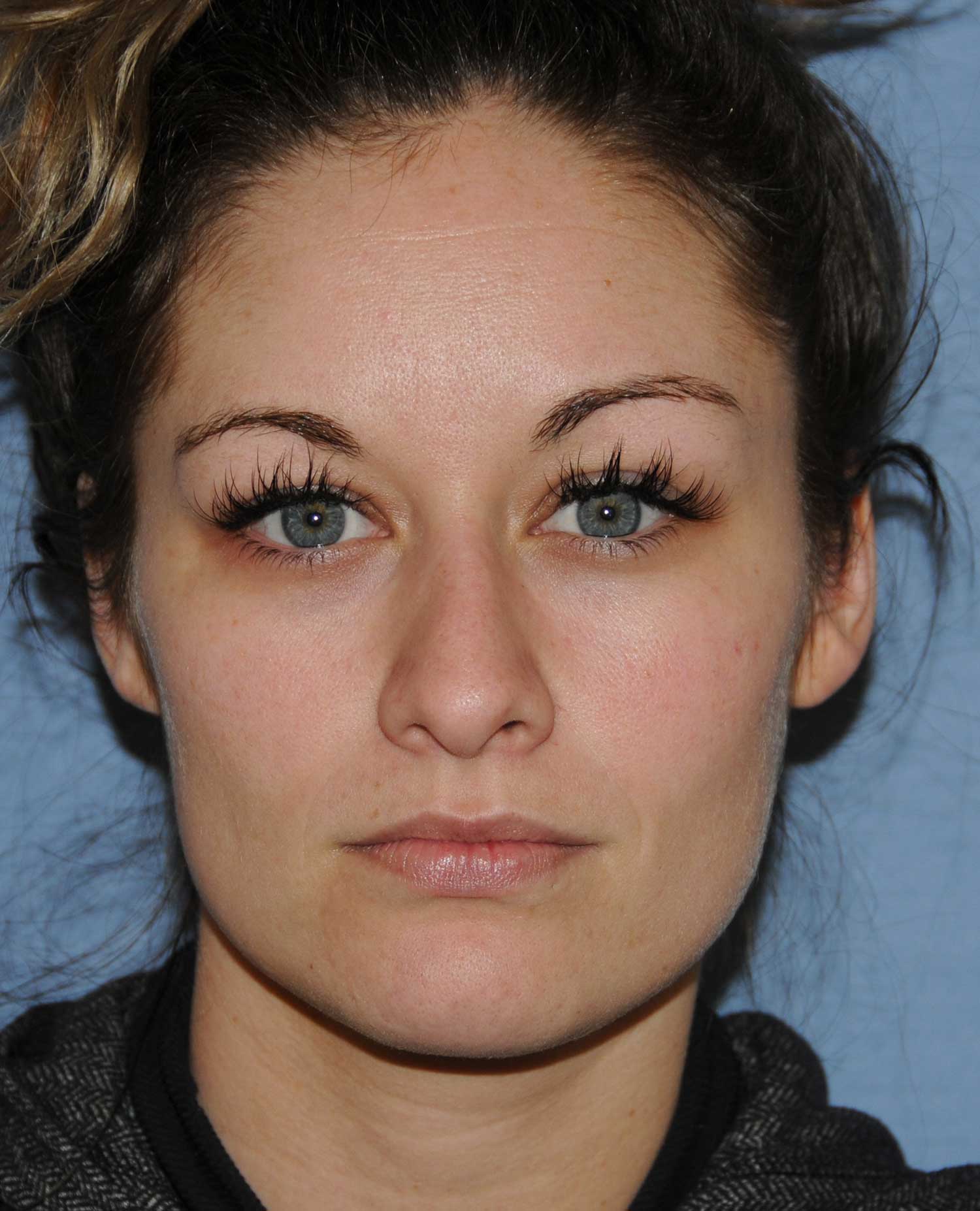
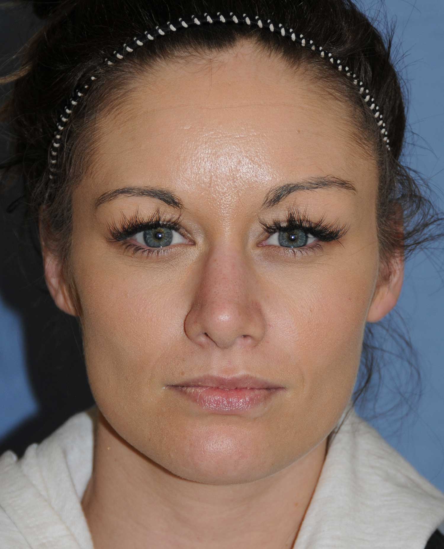
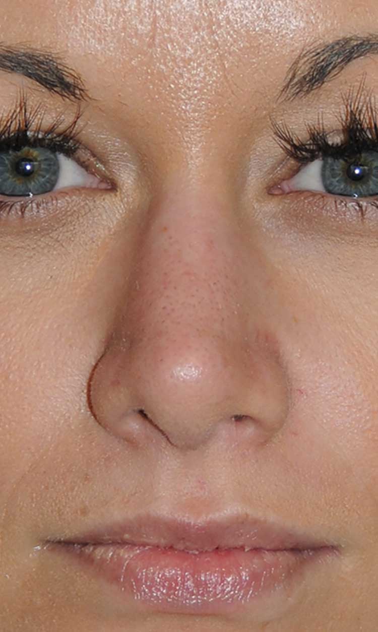
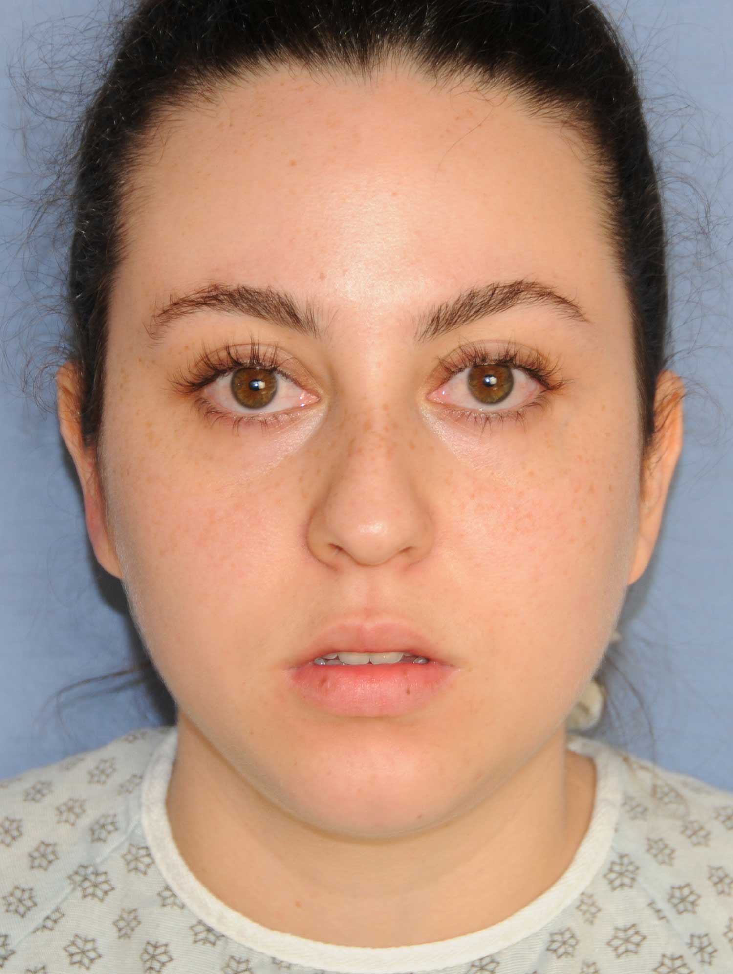
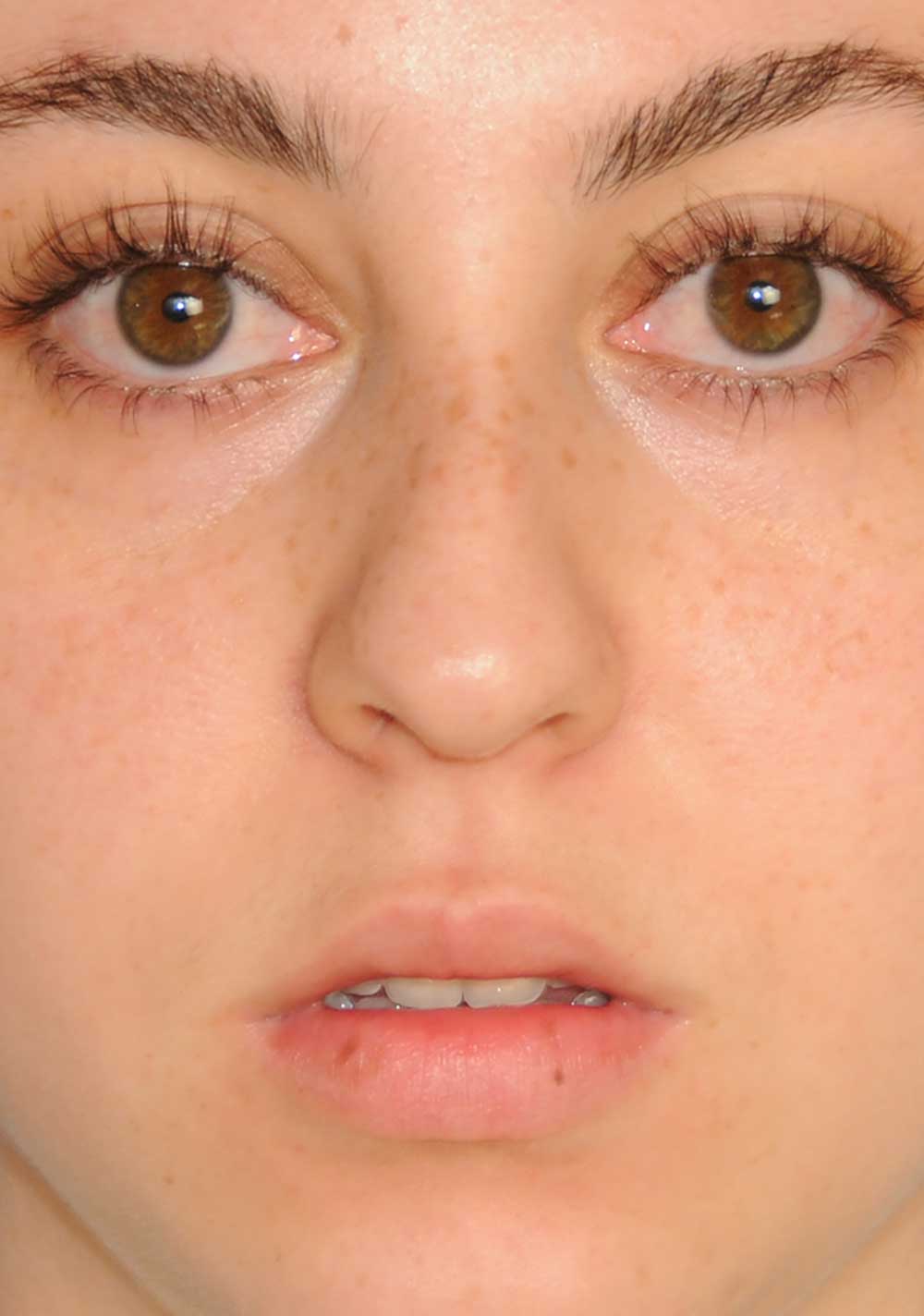
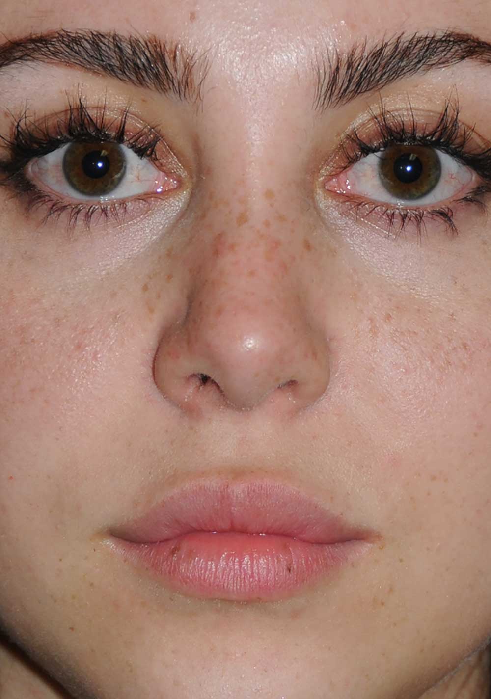
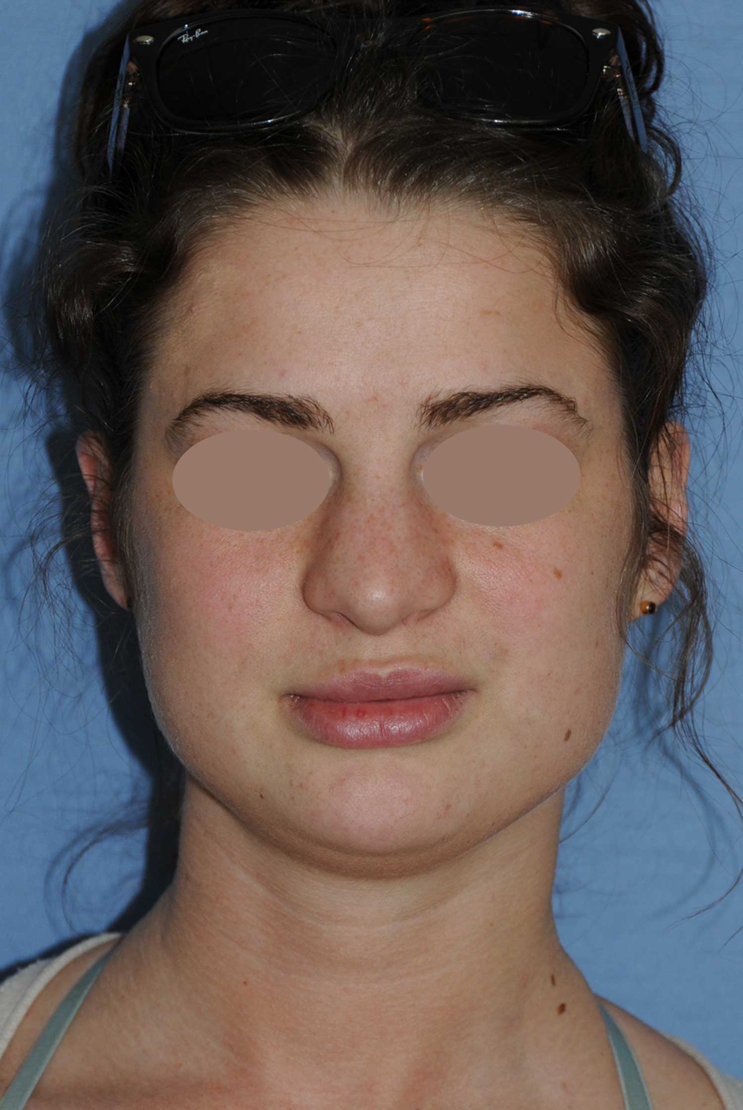
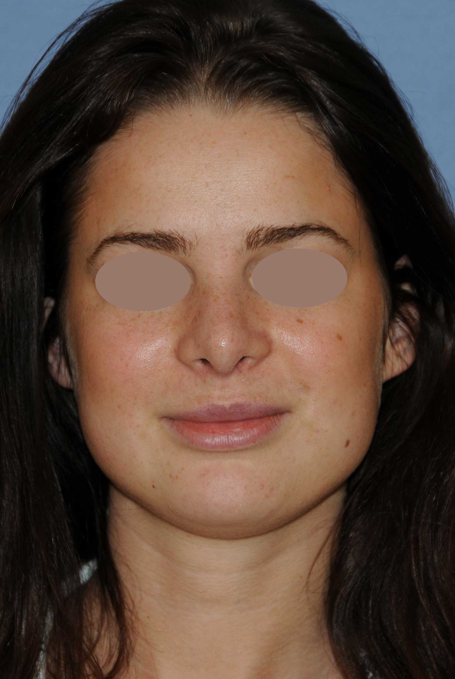
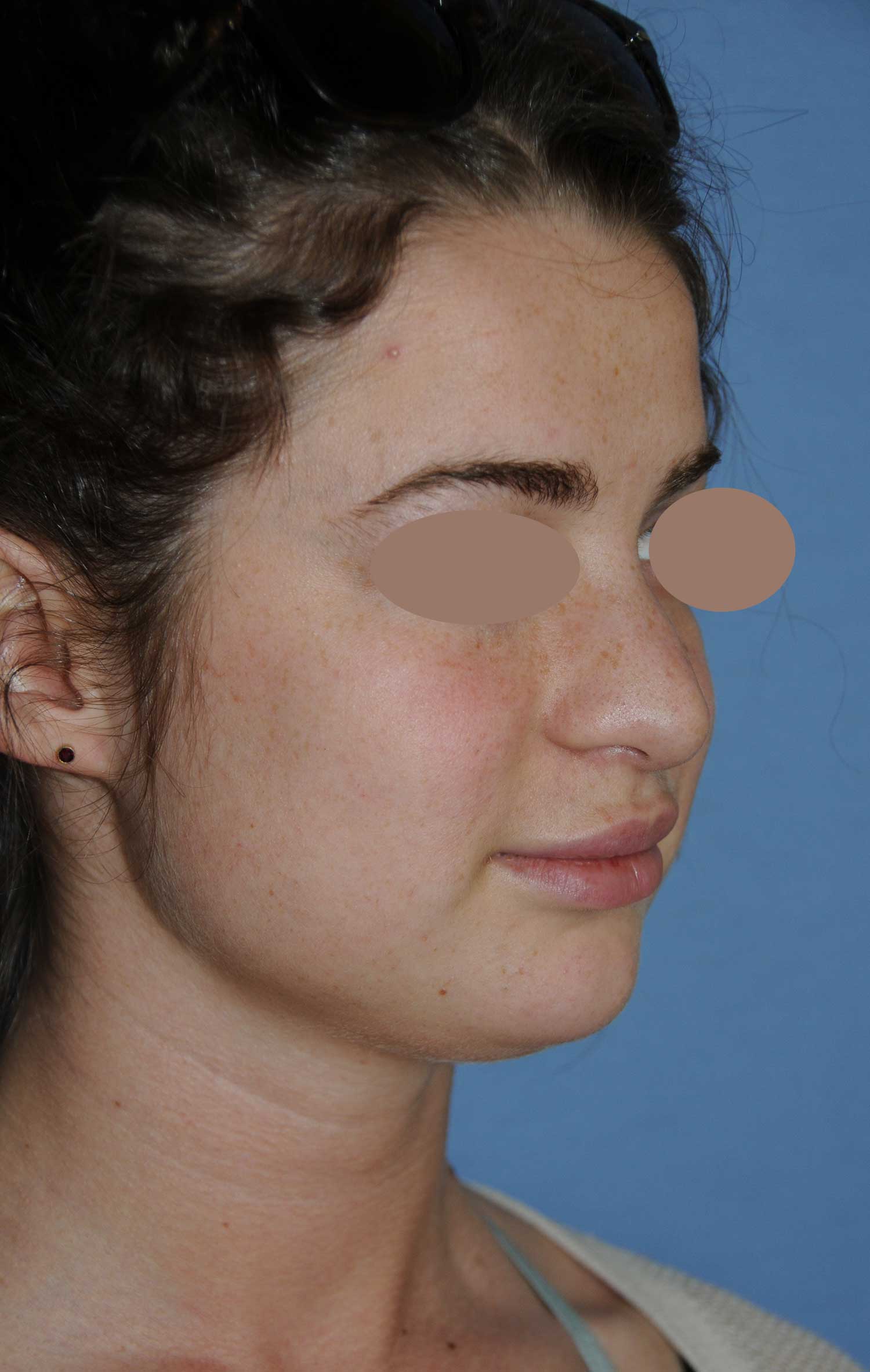
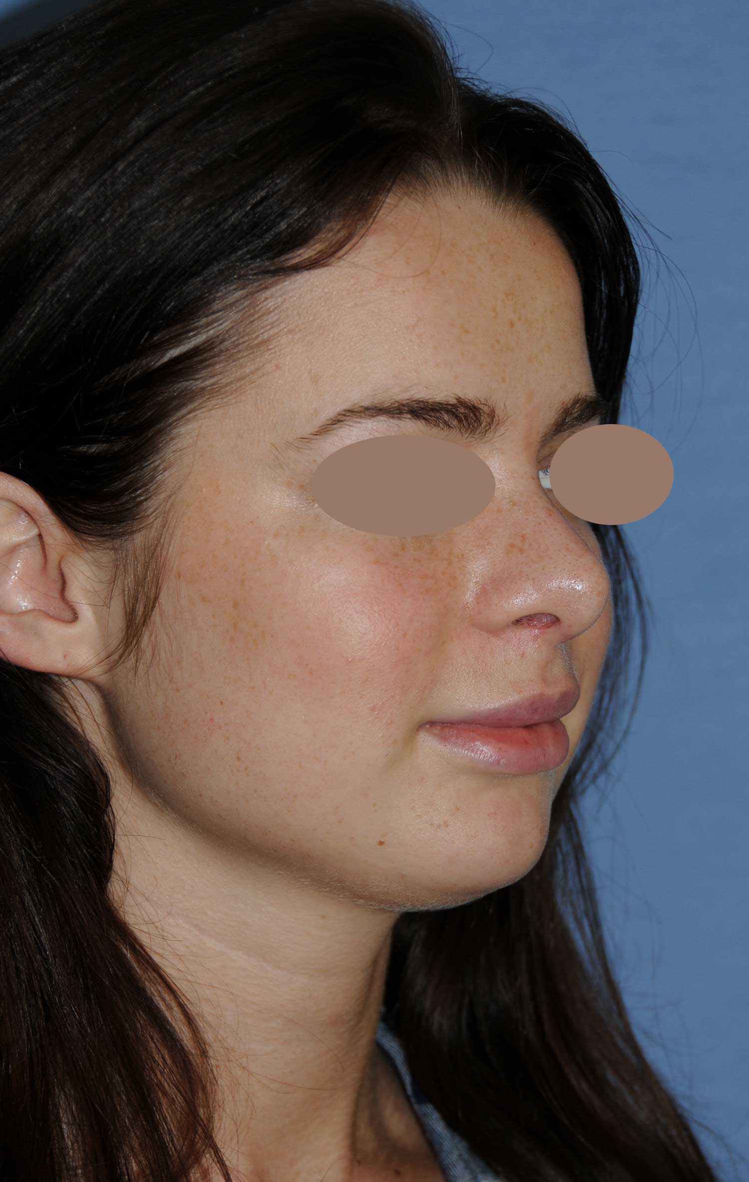
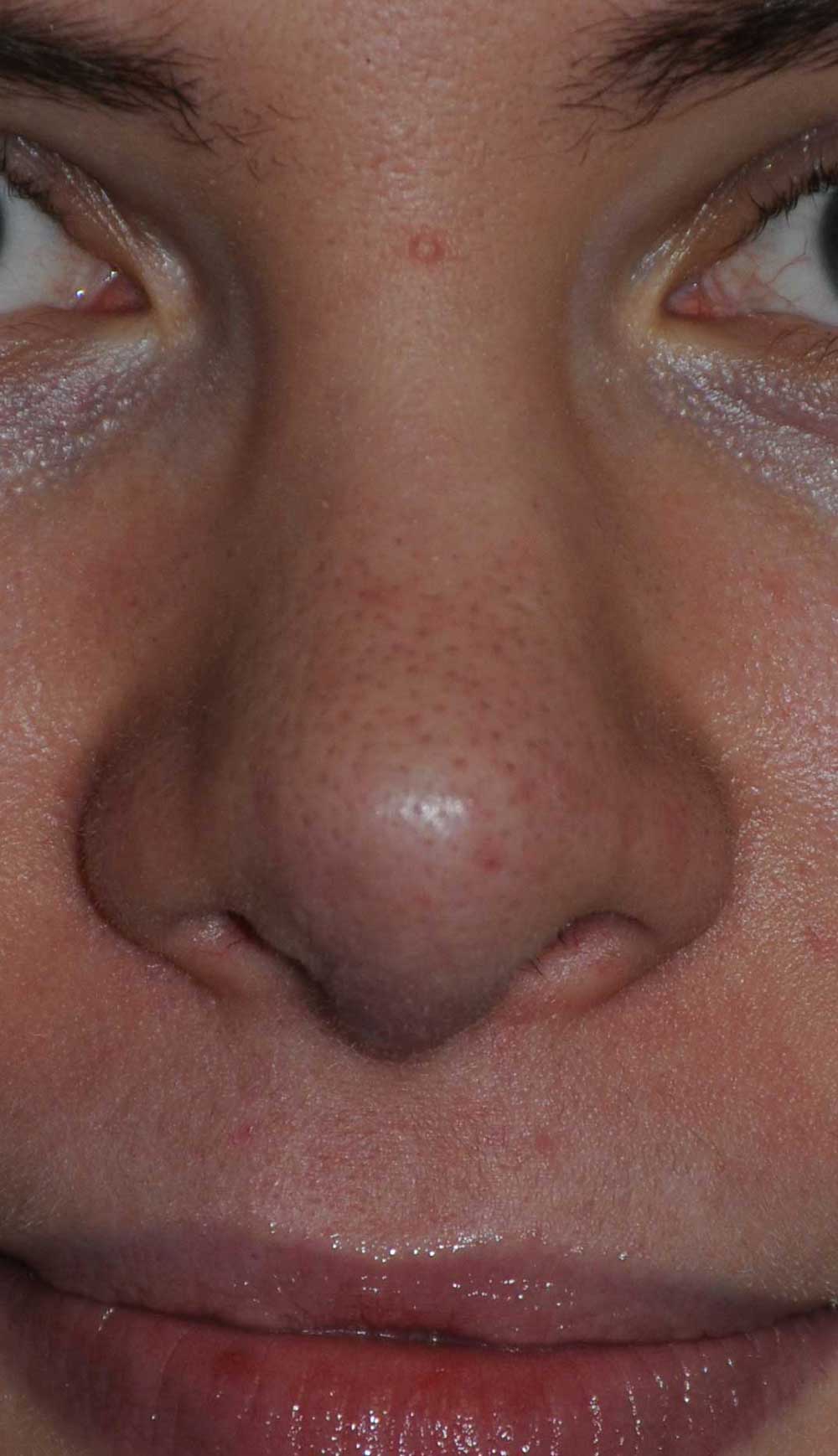
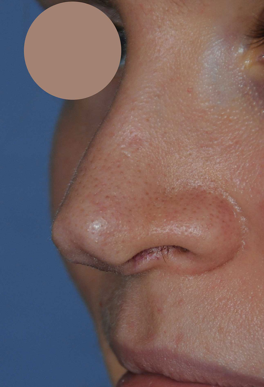
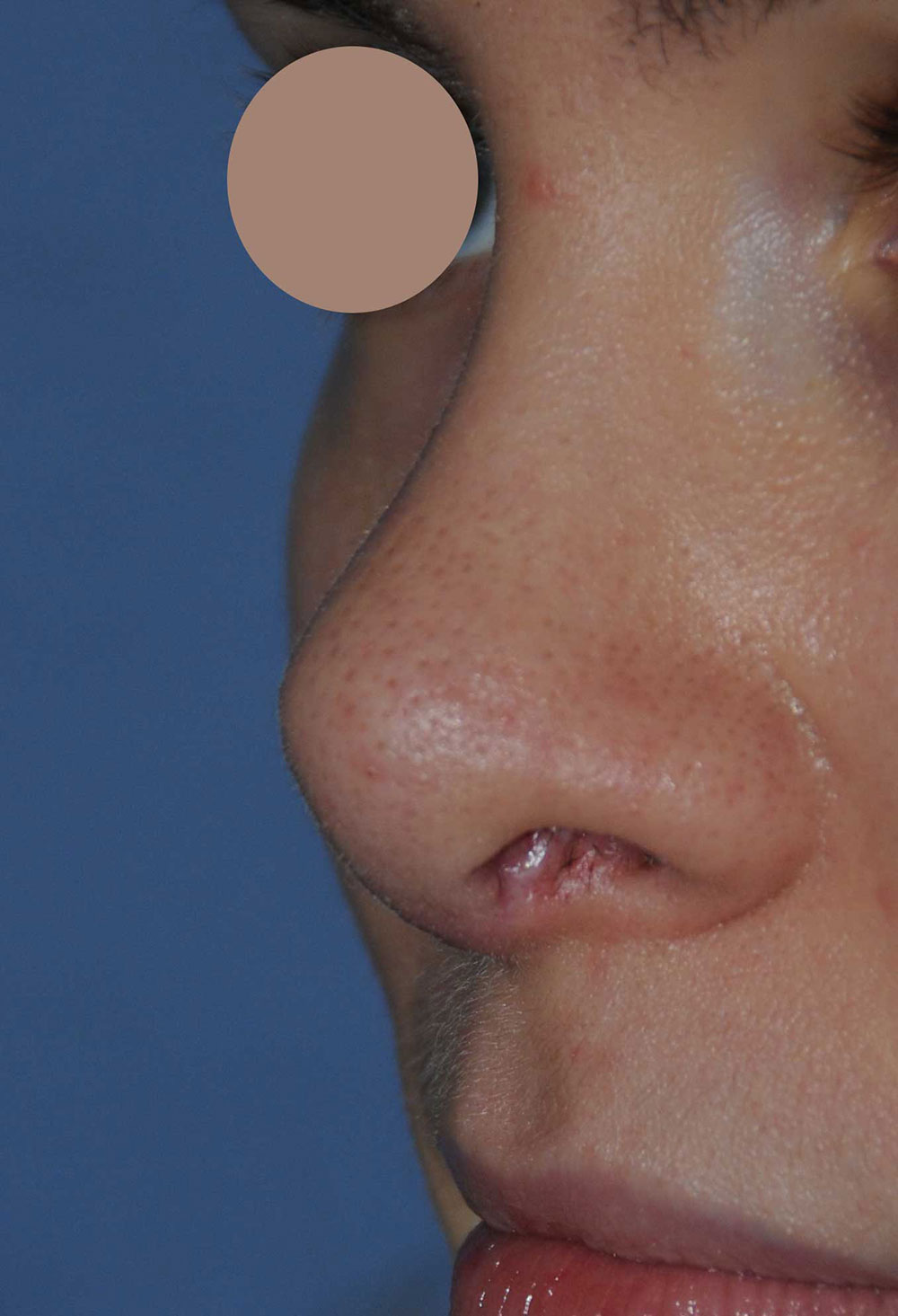
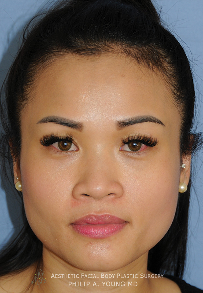
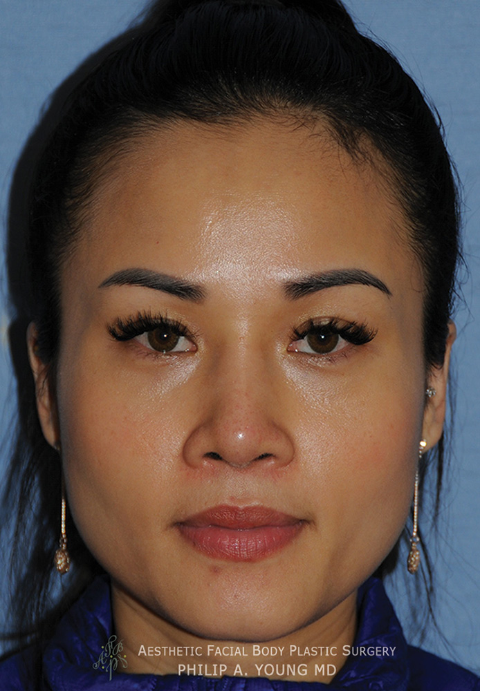
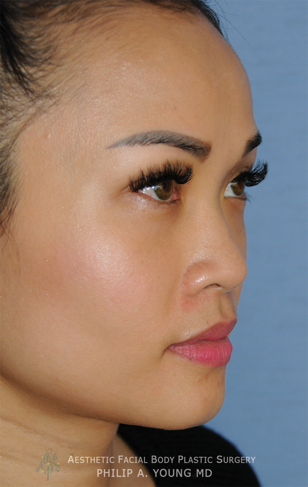
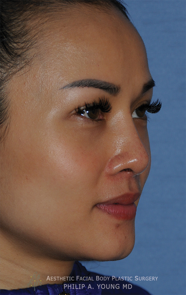
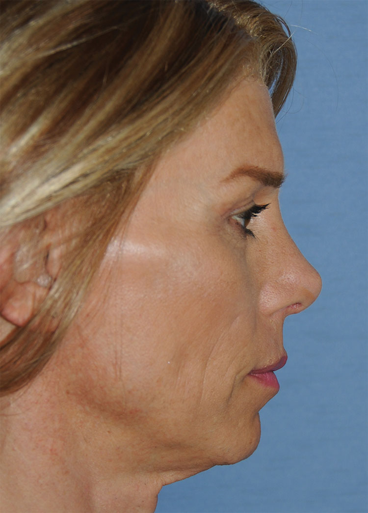
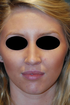
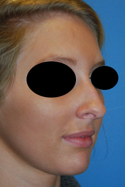
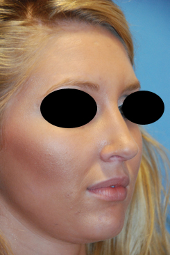
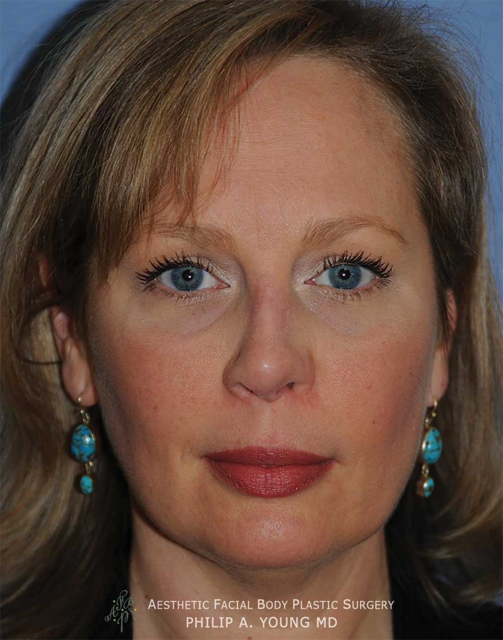
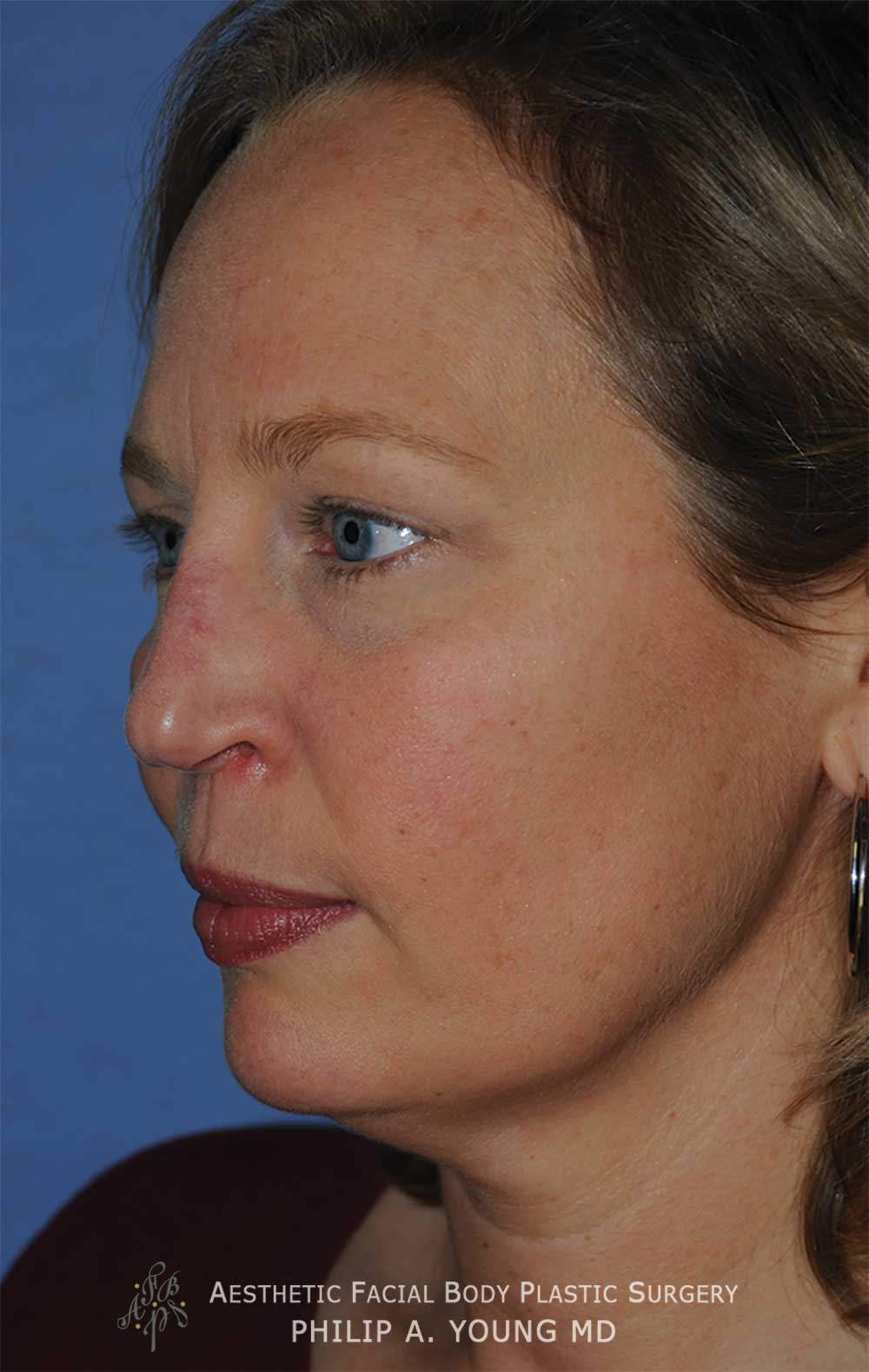
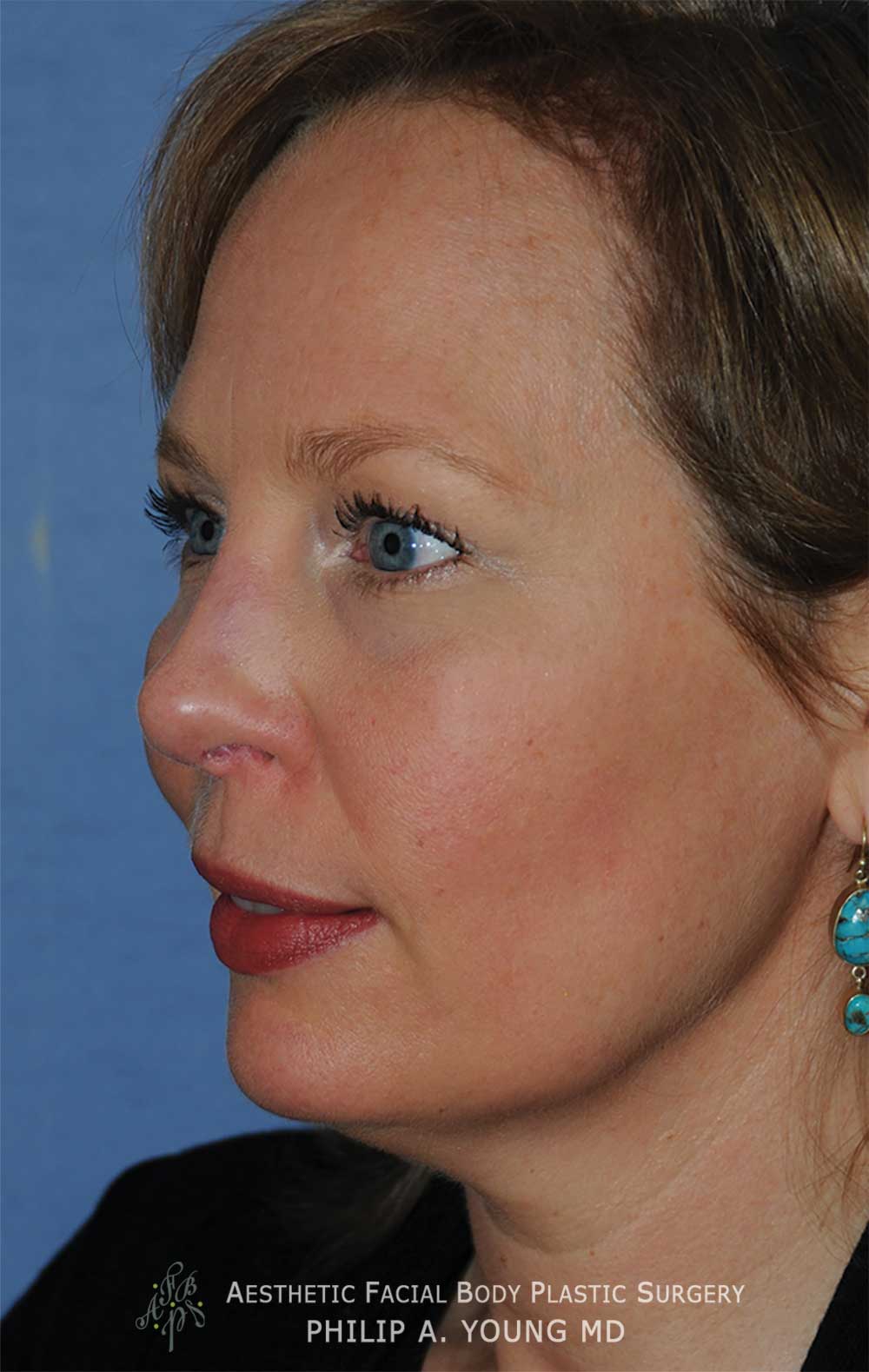
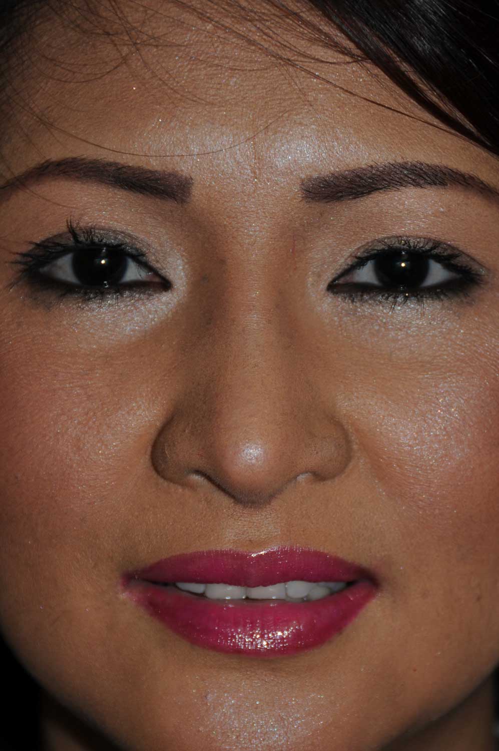
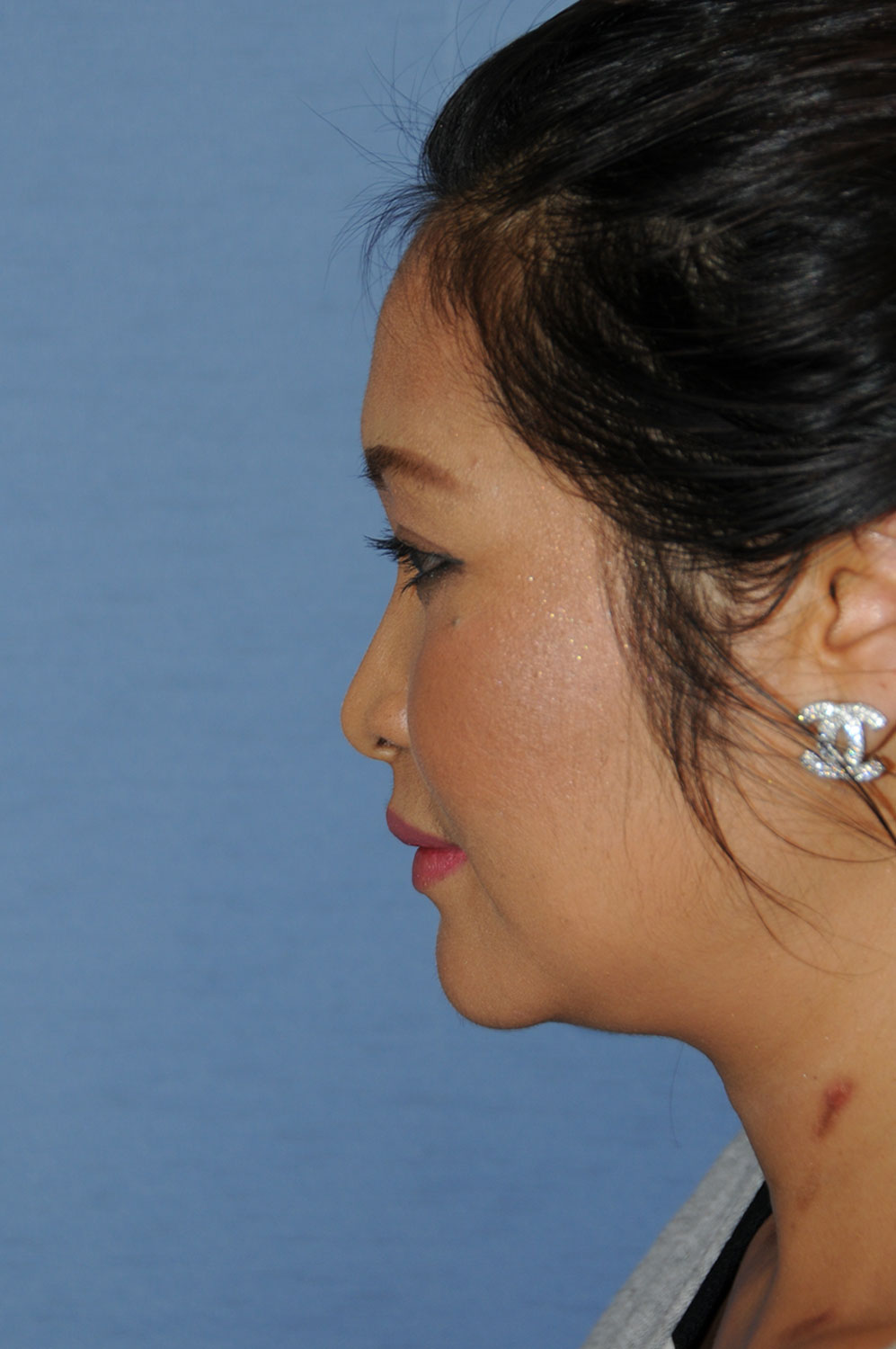
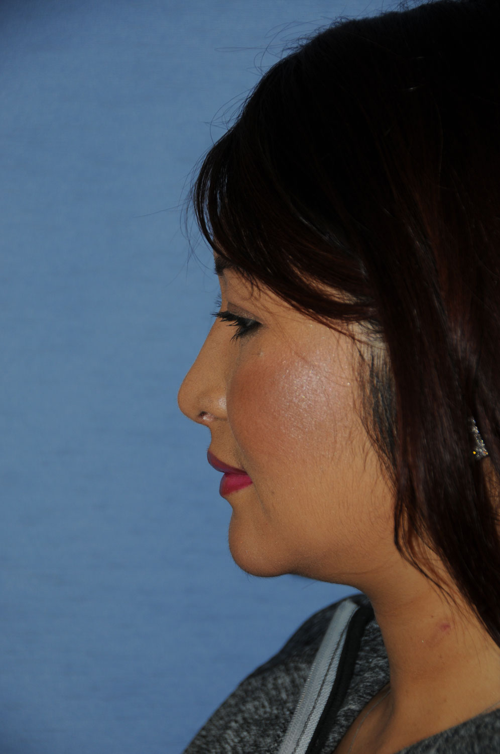
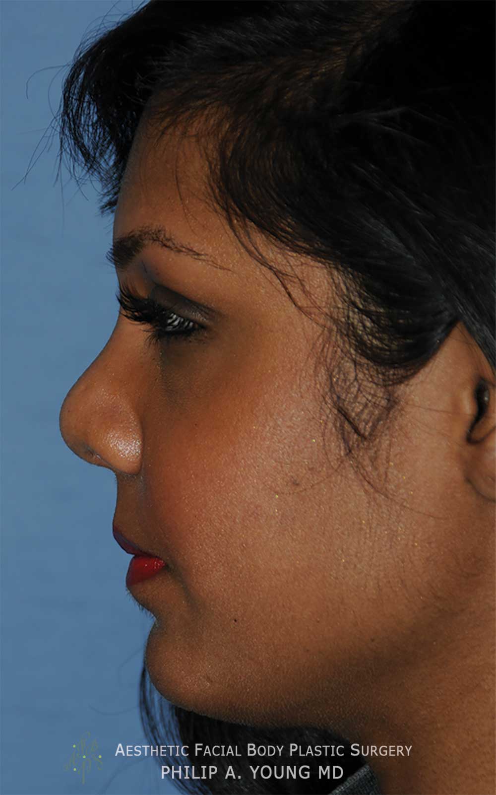
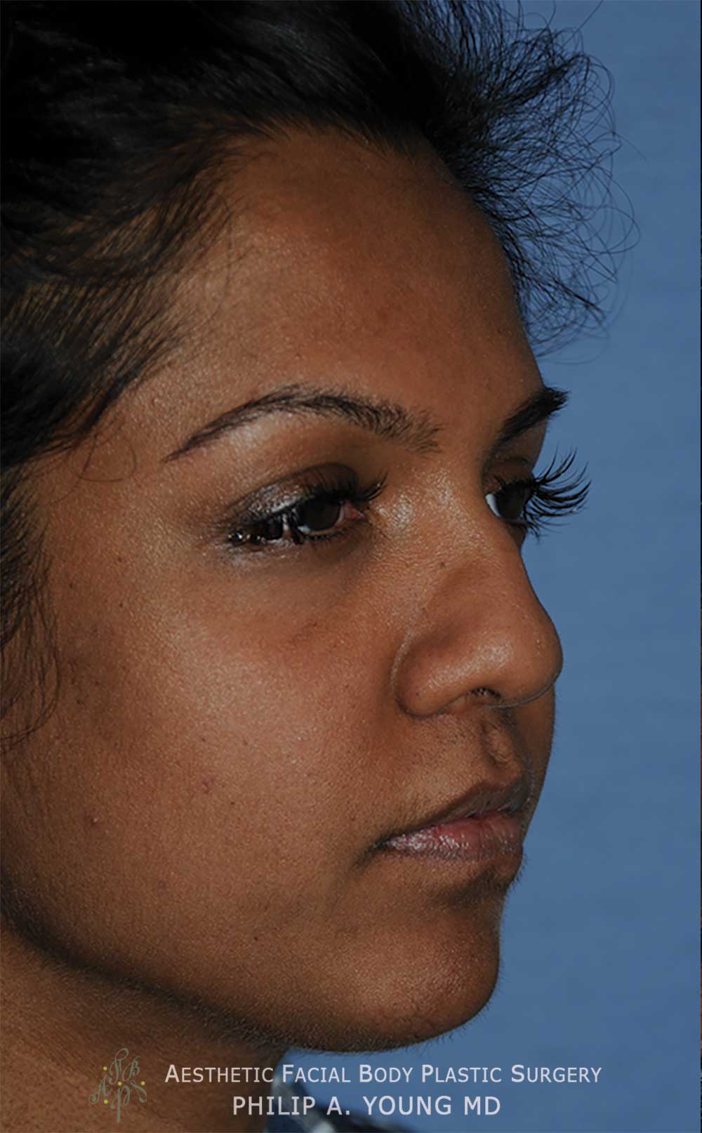
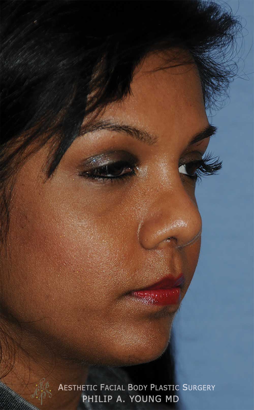
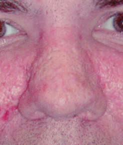
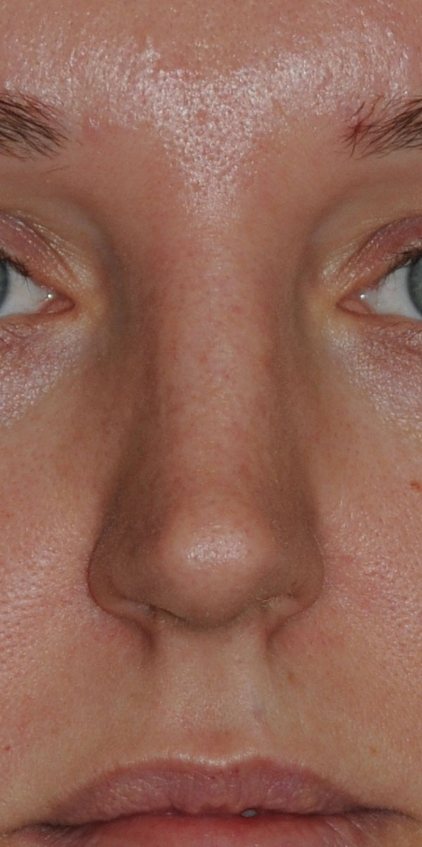
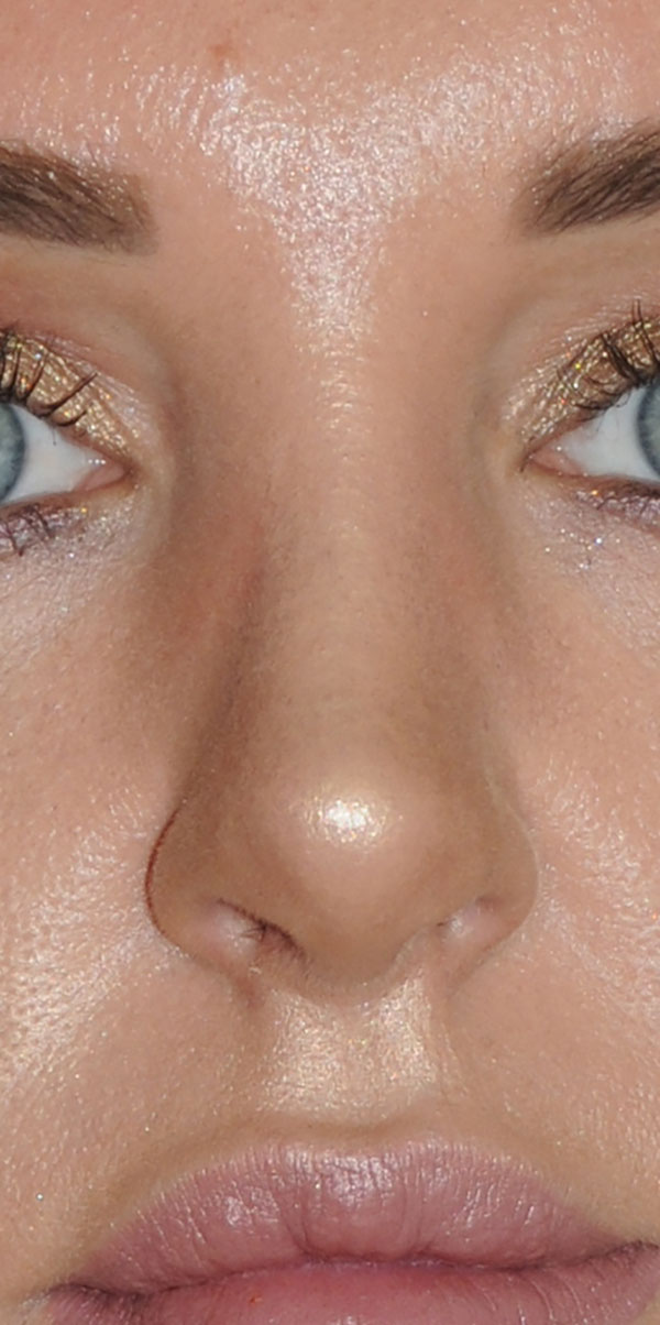
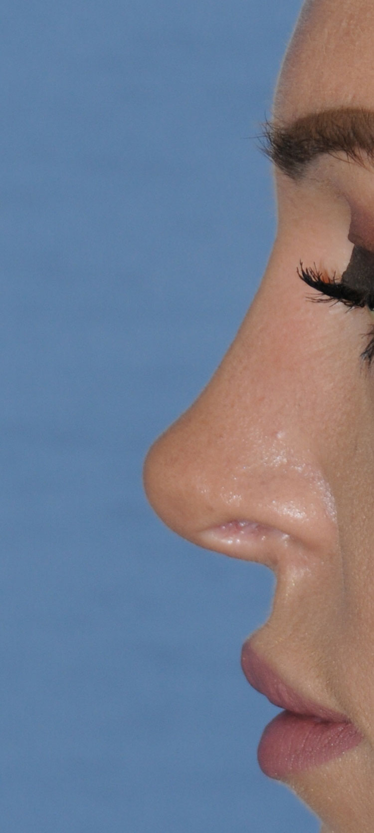
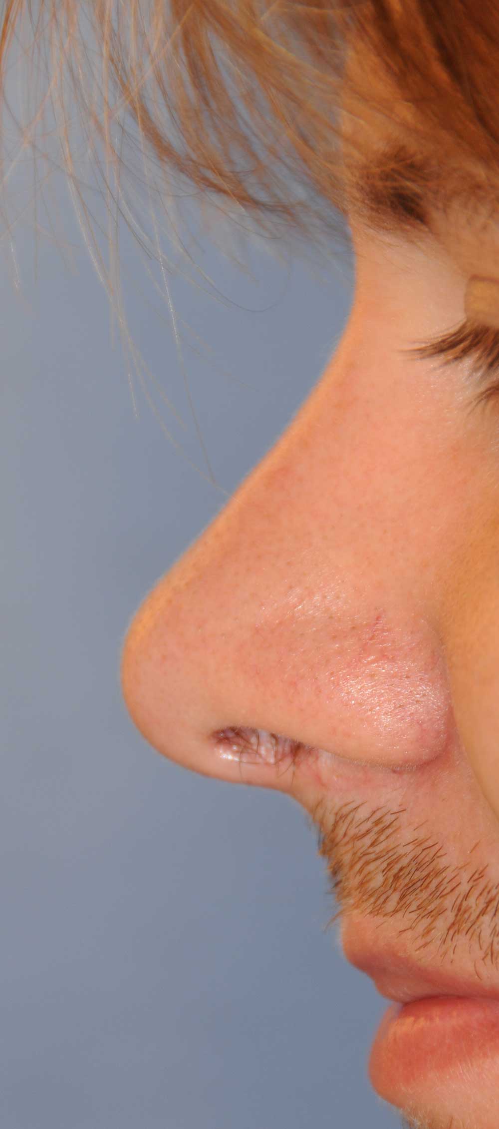
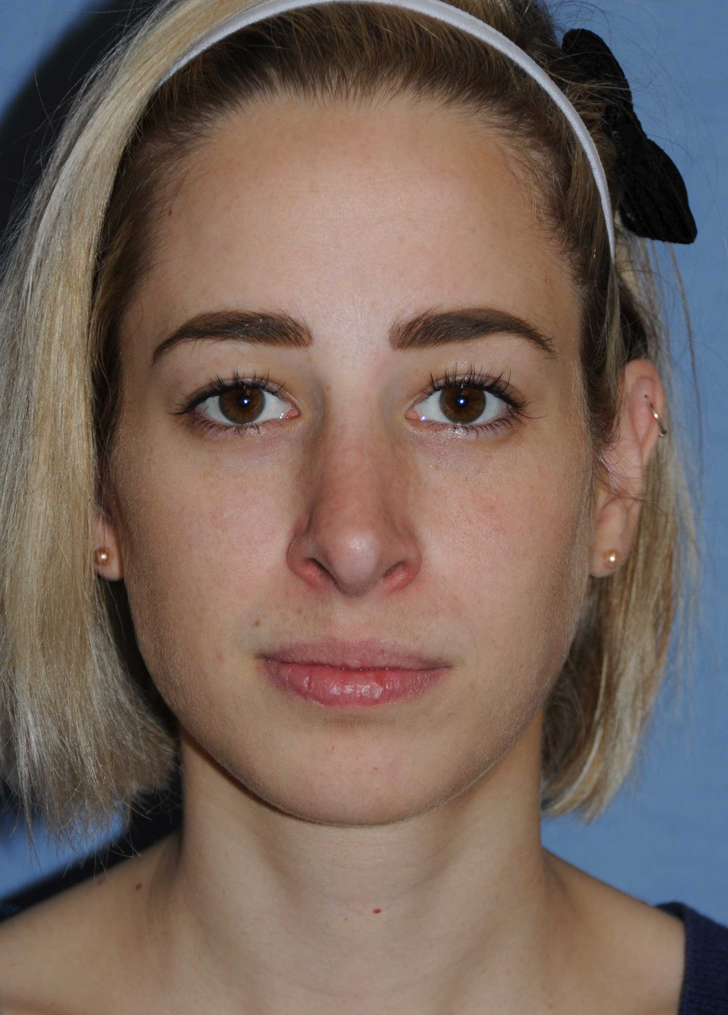
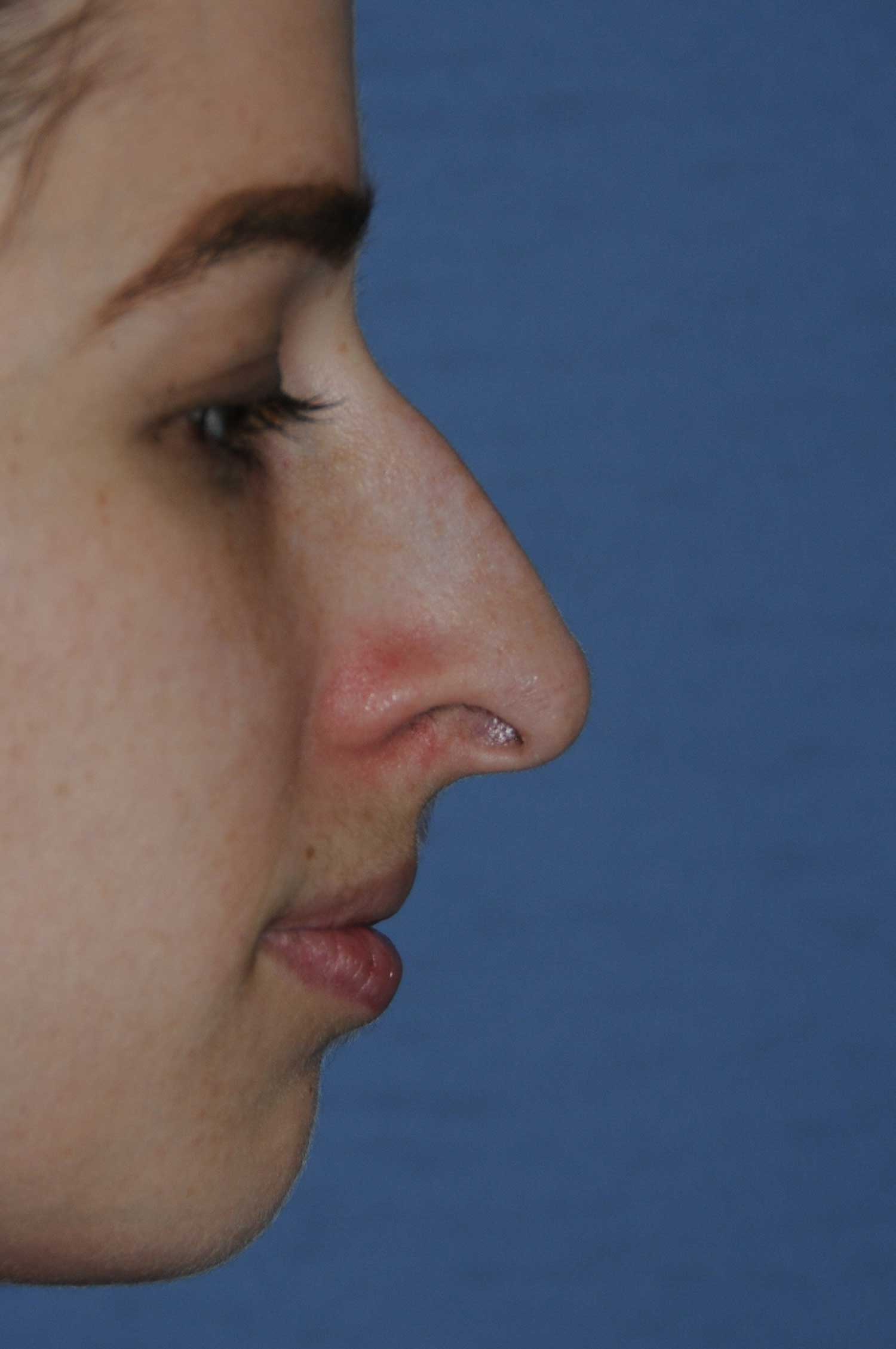
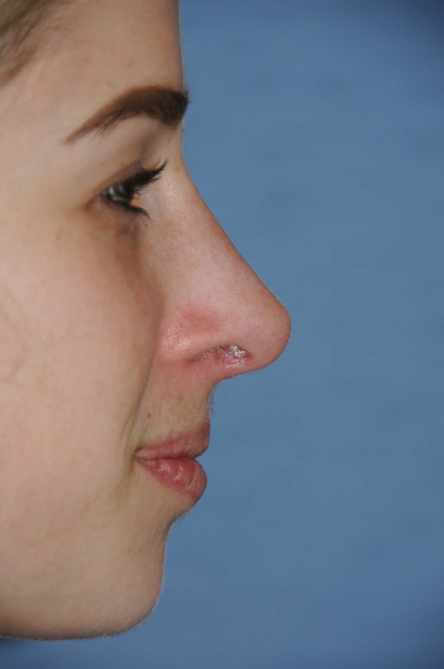
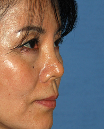
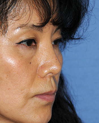
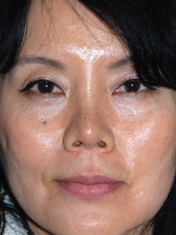
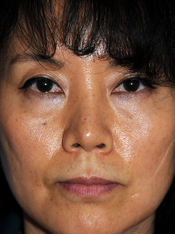
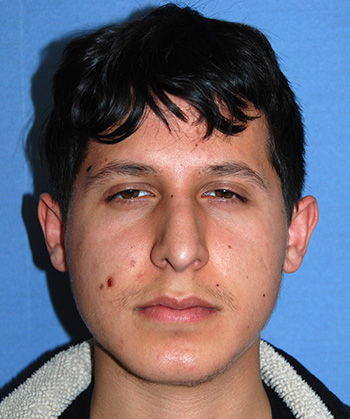
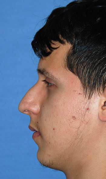
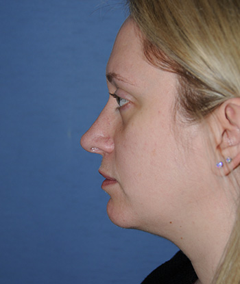
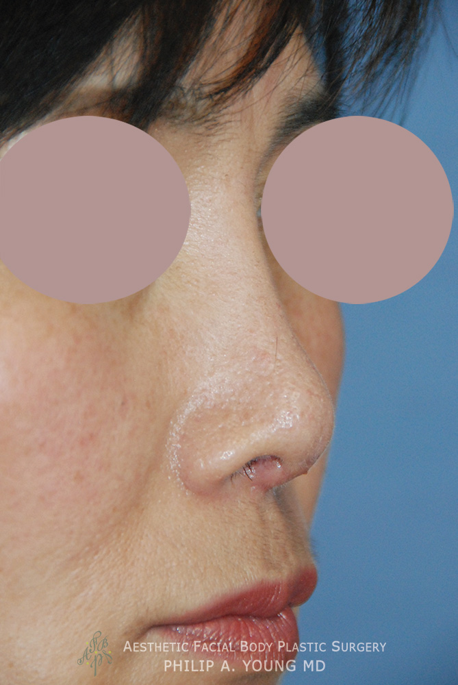
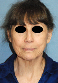
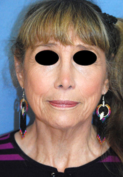
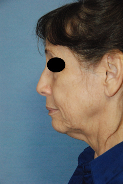
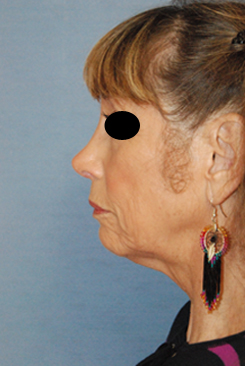
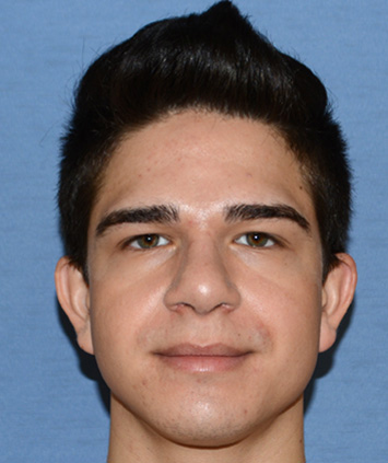
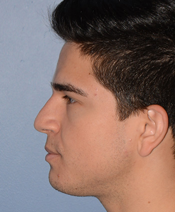
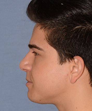
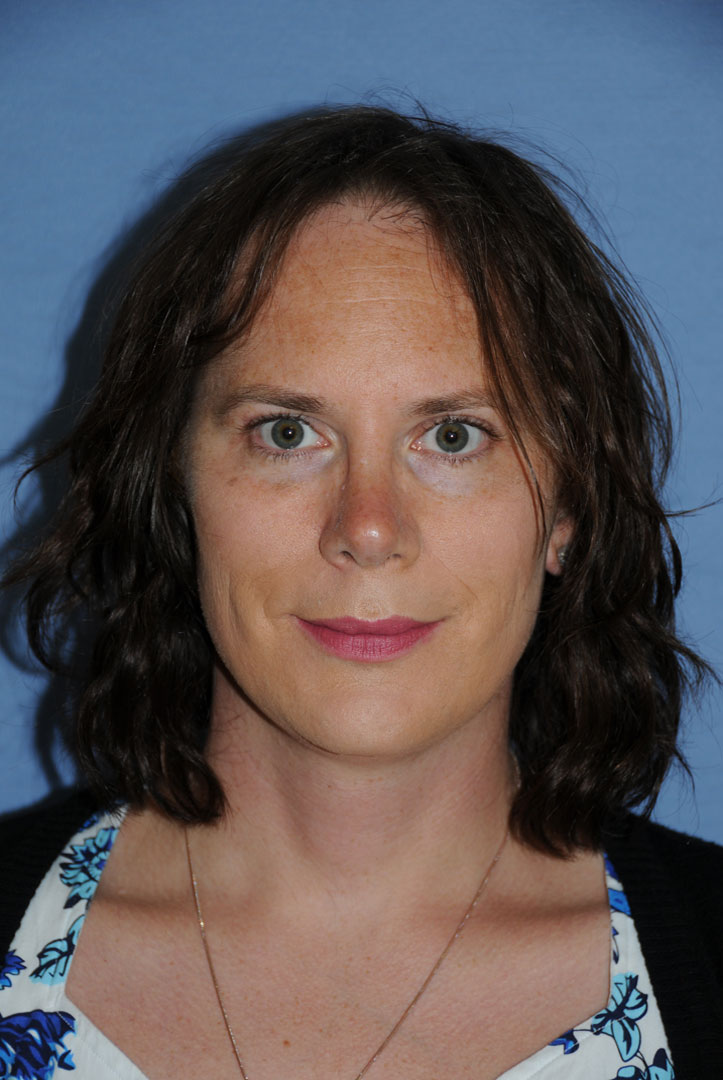
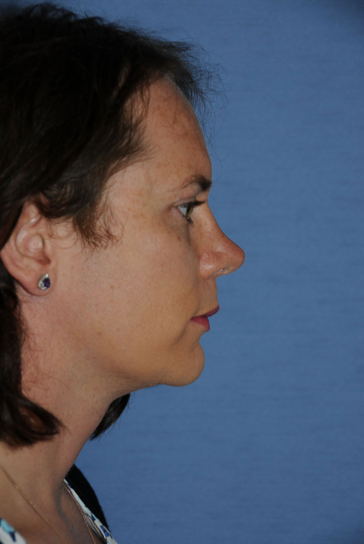
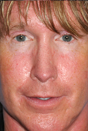
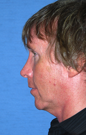
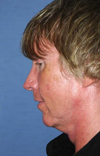
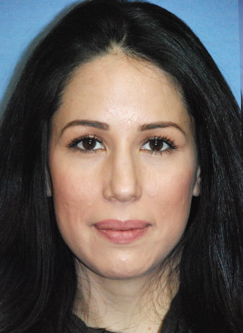
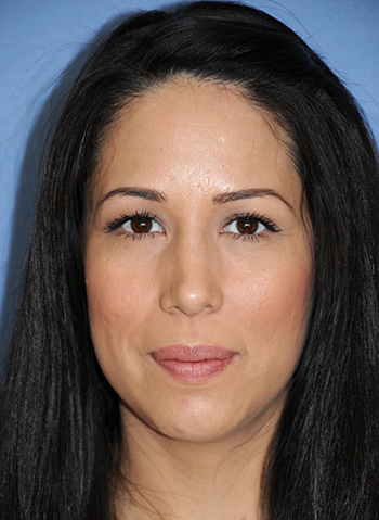
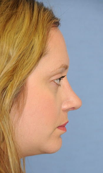
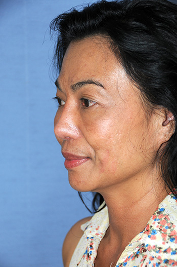
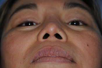
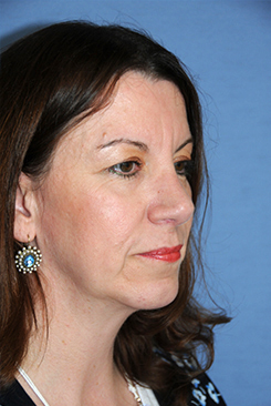
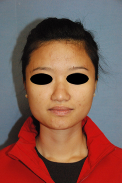
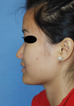
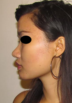
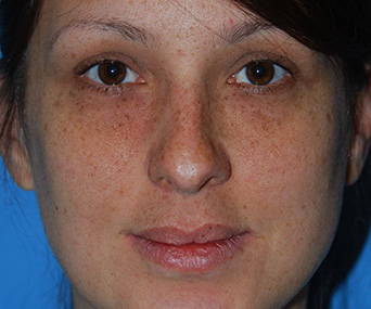
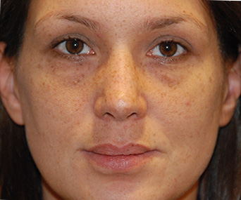
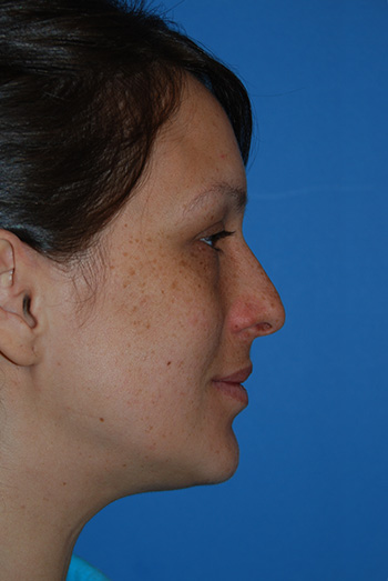
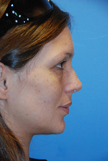
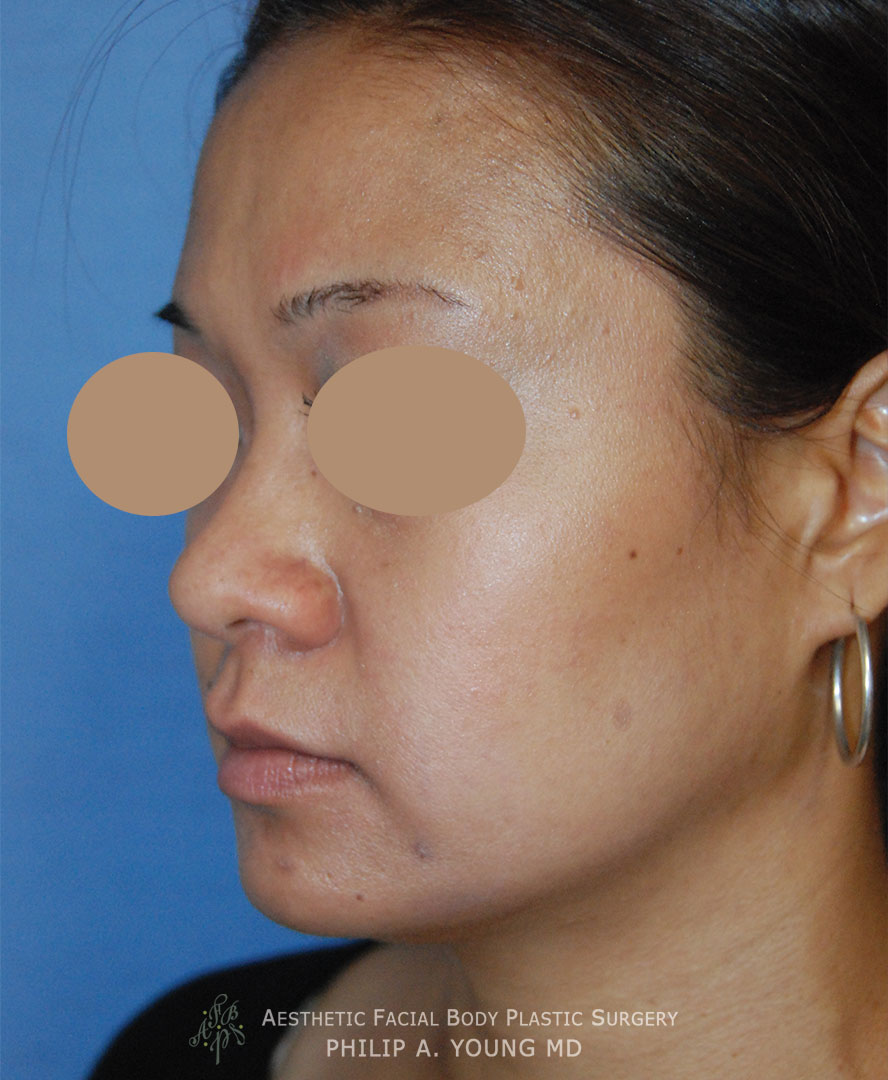
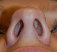
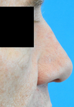
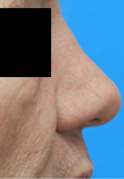
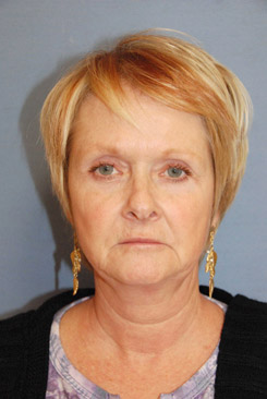
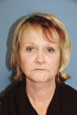
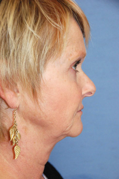
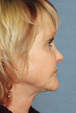
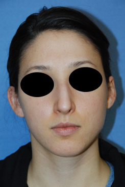
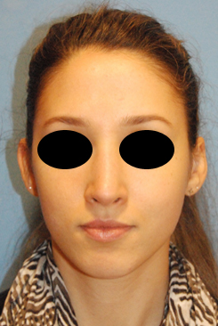
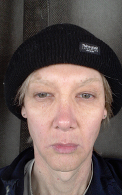
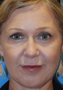
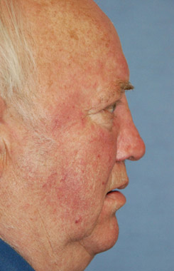
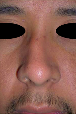
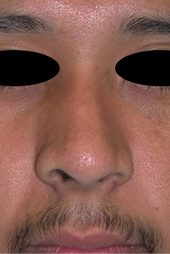
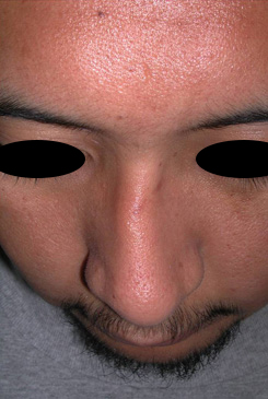
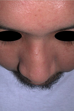
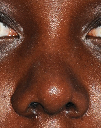
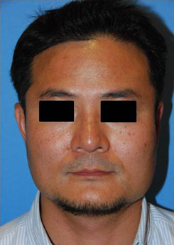
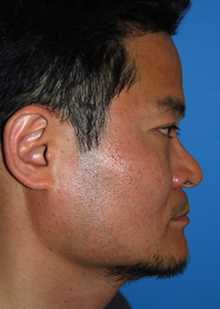
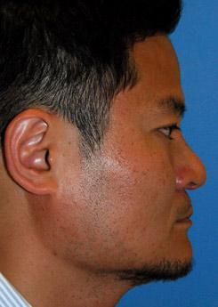
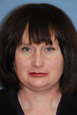
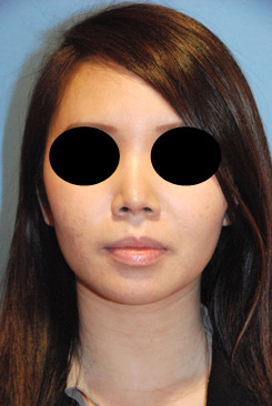
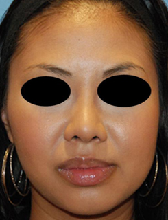
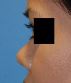
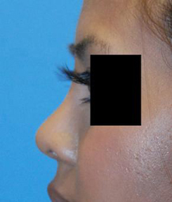
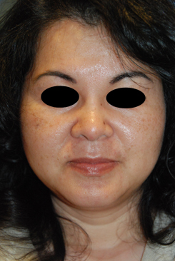
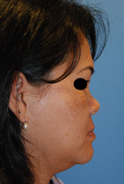
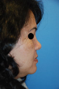
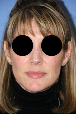
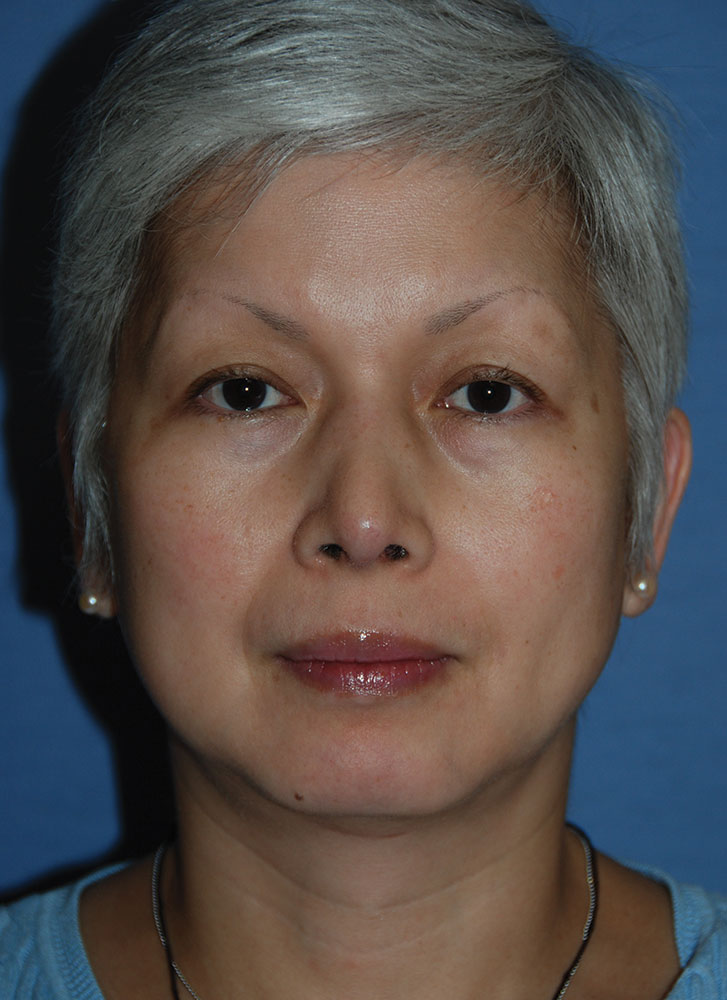
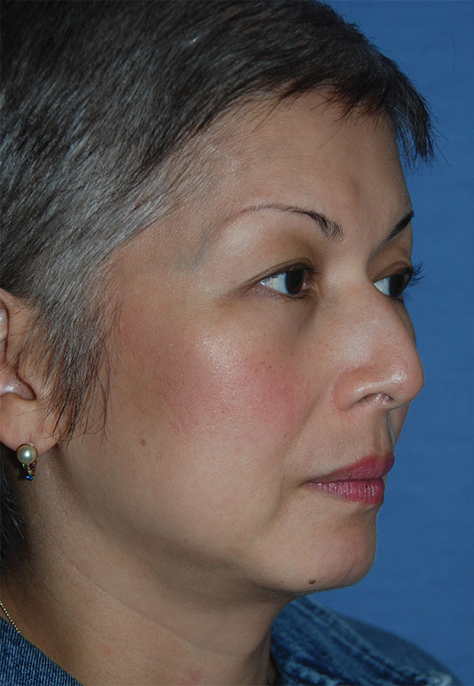
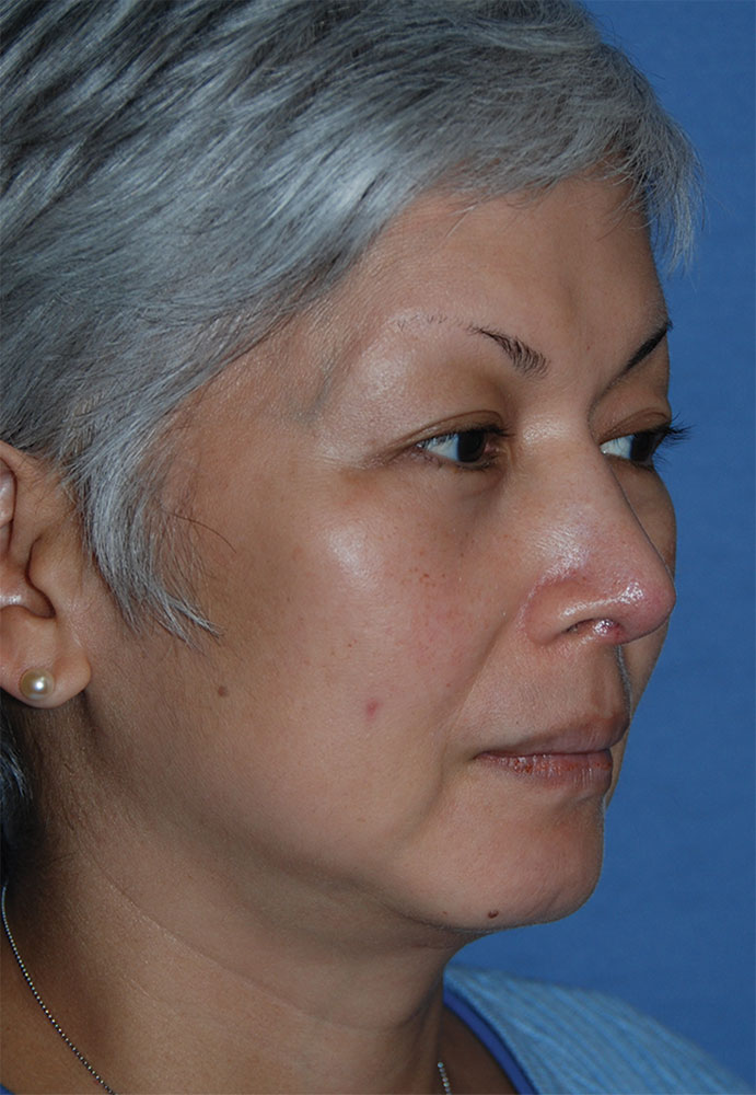
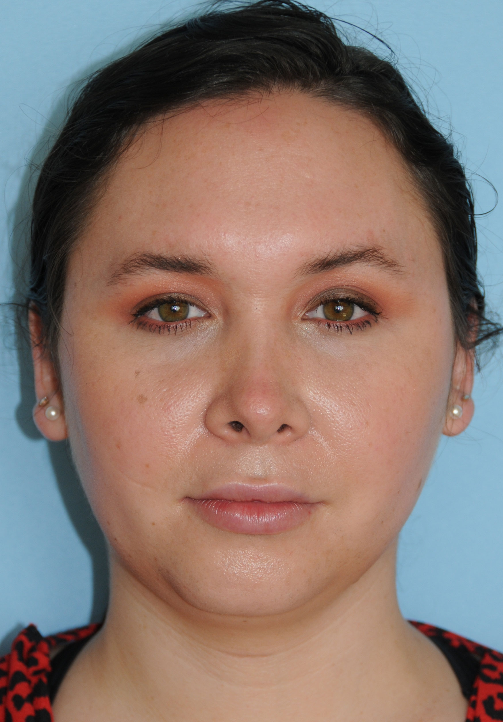
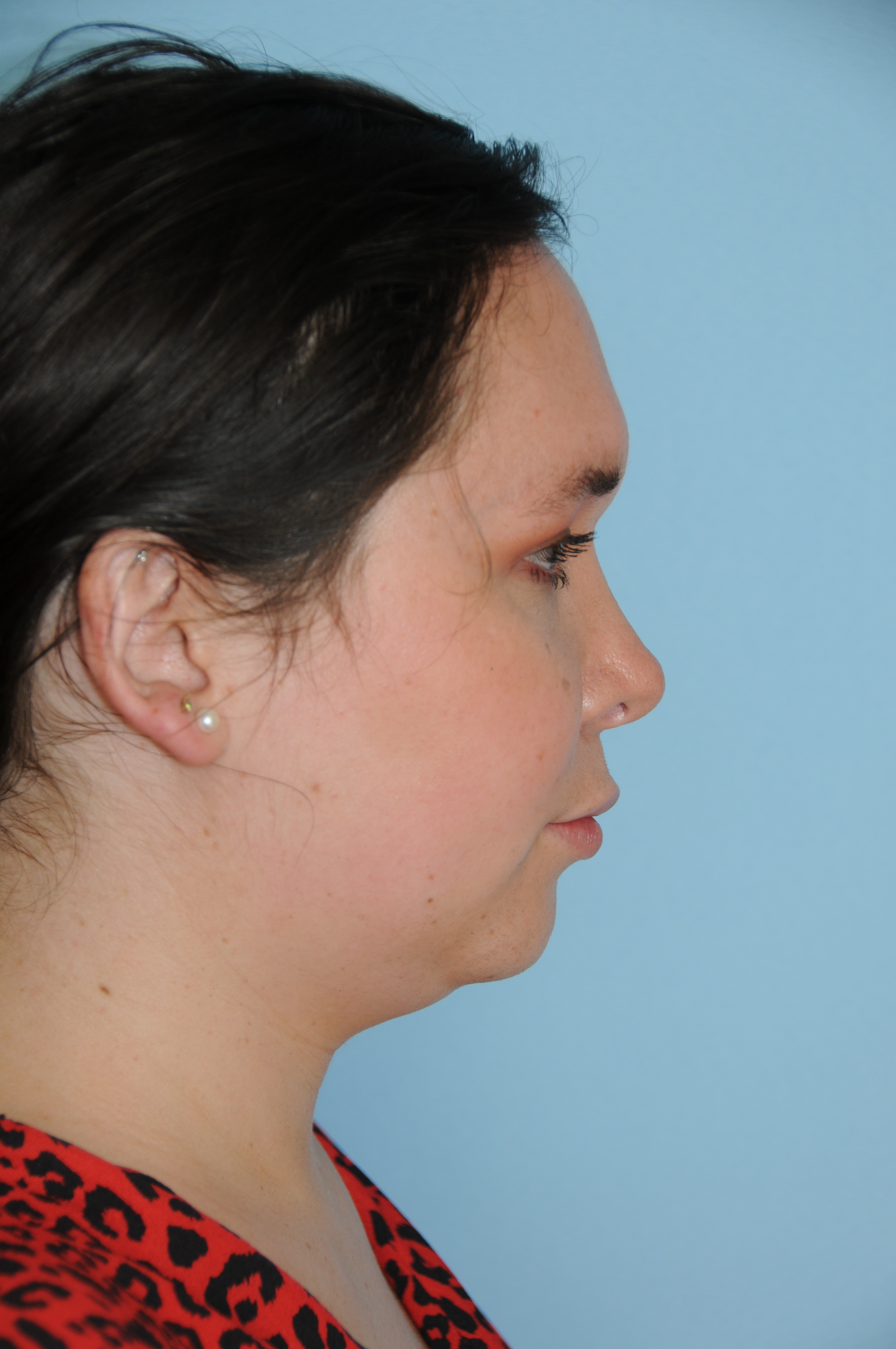
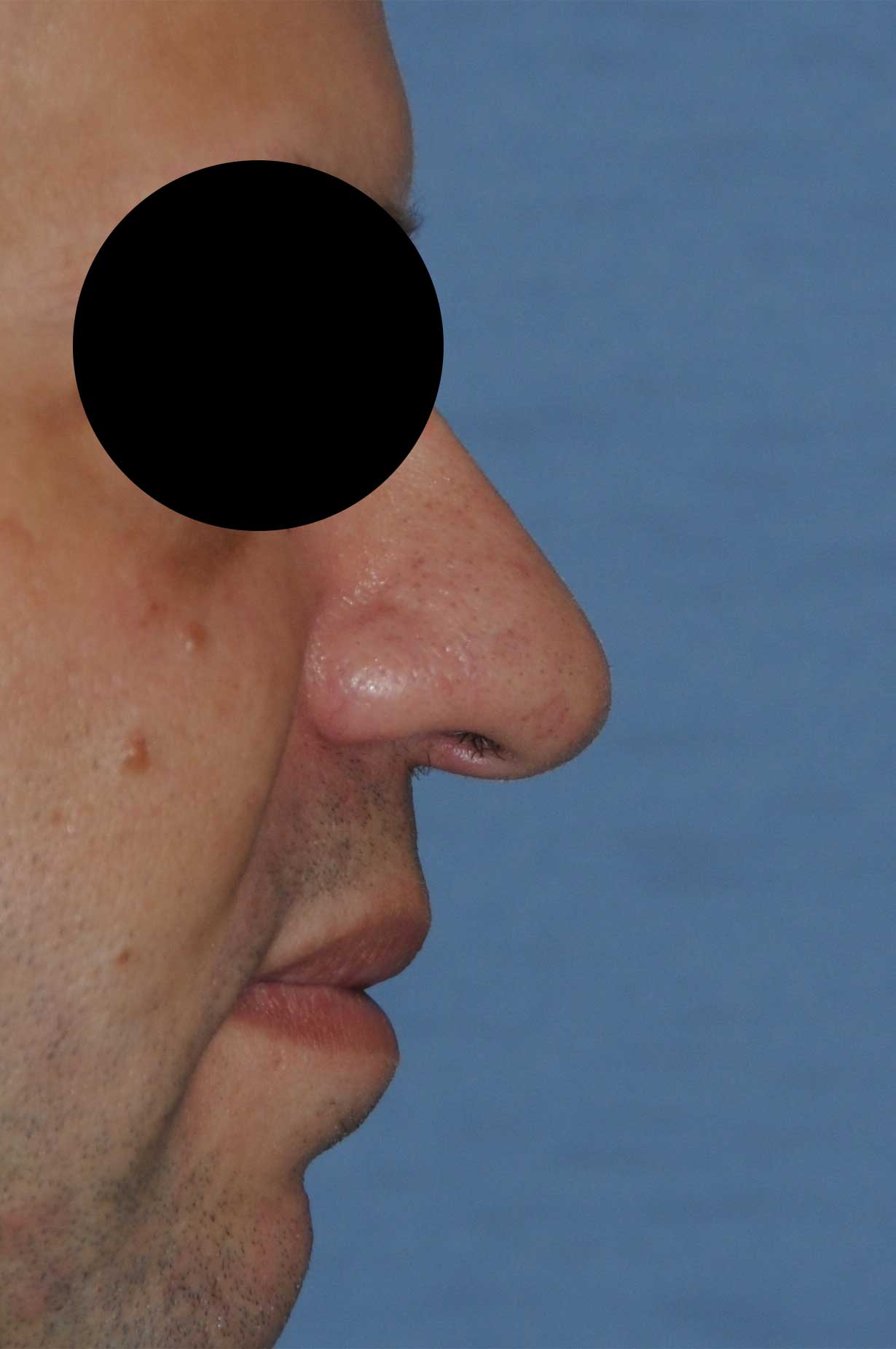
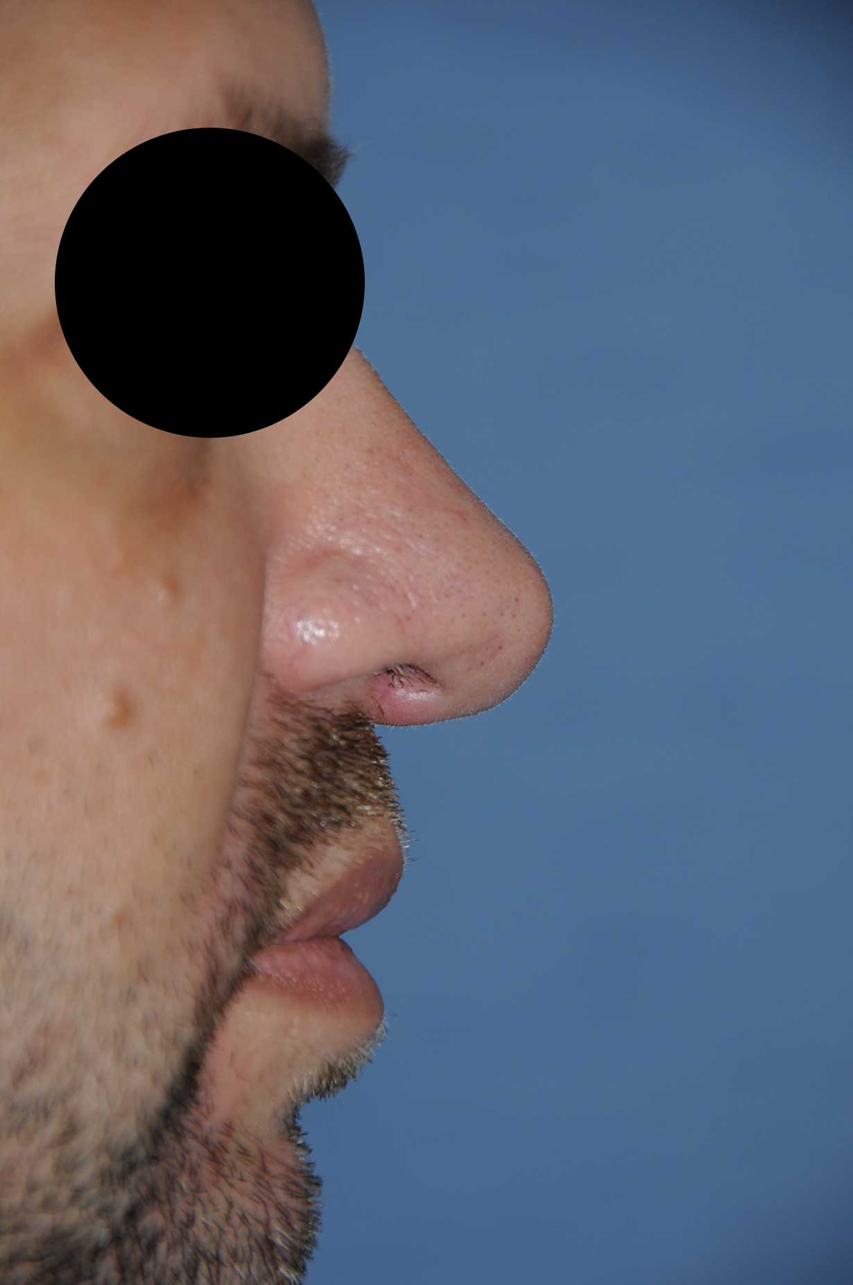
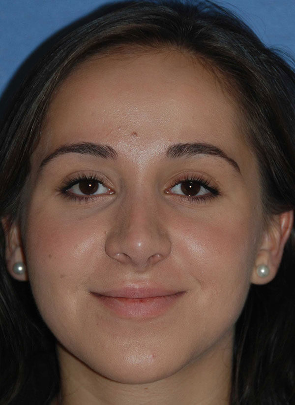
This page was created by Seattle Rhinoplasty Expert Dr. Philip Young to help you understand the terms that will be used for your Rhinoplasty procedure. It will help facilitate your understanding from your Rhinoplasty Consultation to Beautiful Results. Below is a picture of one of our happy Rhinoplasty patients. We will show you her picture before defining some anatomy terms of the nose:
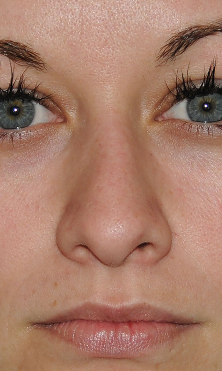
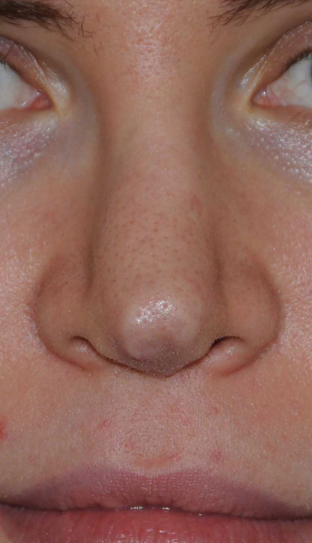
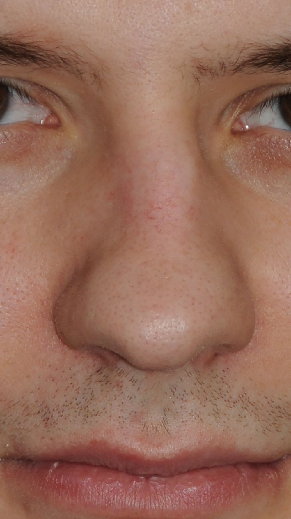
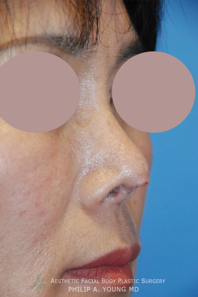
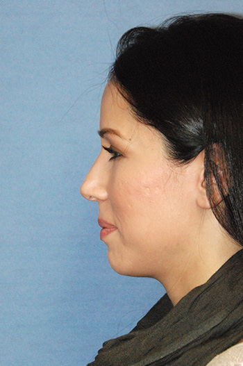
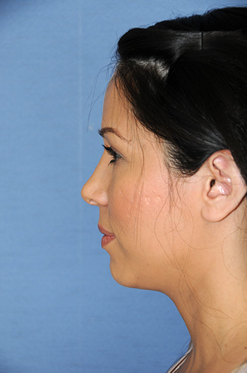
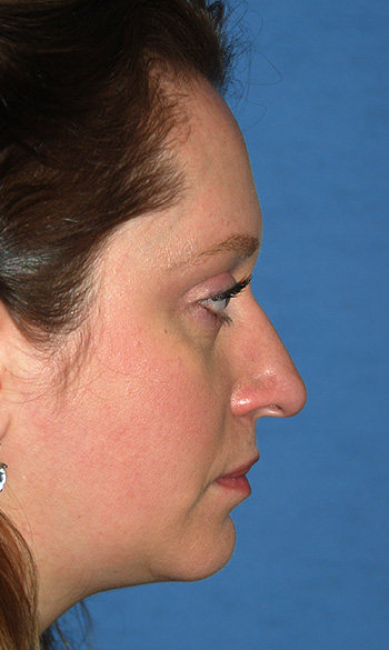
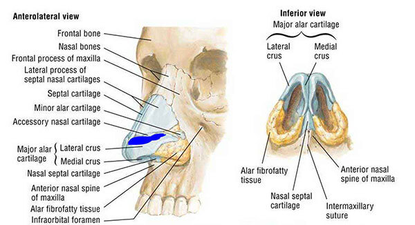
Columella: Is the structure between the nostrils that is made of medial crura, the cartilage of the lower third of the nose. The lower lateral cartilages which will be defined later is the cartilage that makes up the tip and lower third. It is made up of 3 parts: the medial crura | segment, the intermediate or middle crura | segment, and the lateral crura | segment. The columella is the area that your incision is made in a gull wing type fashion in an open approach.
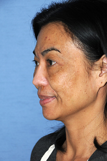
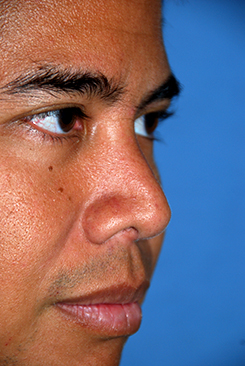
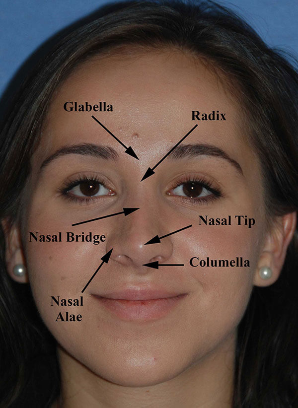
Glabella: This is the prominence made of bone, underlying frontal sinus and soft tissue that lies between the eyebrows
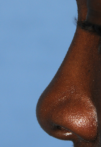
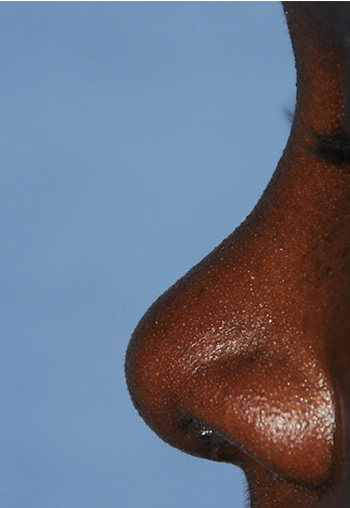
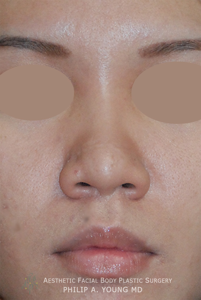
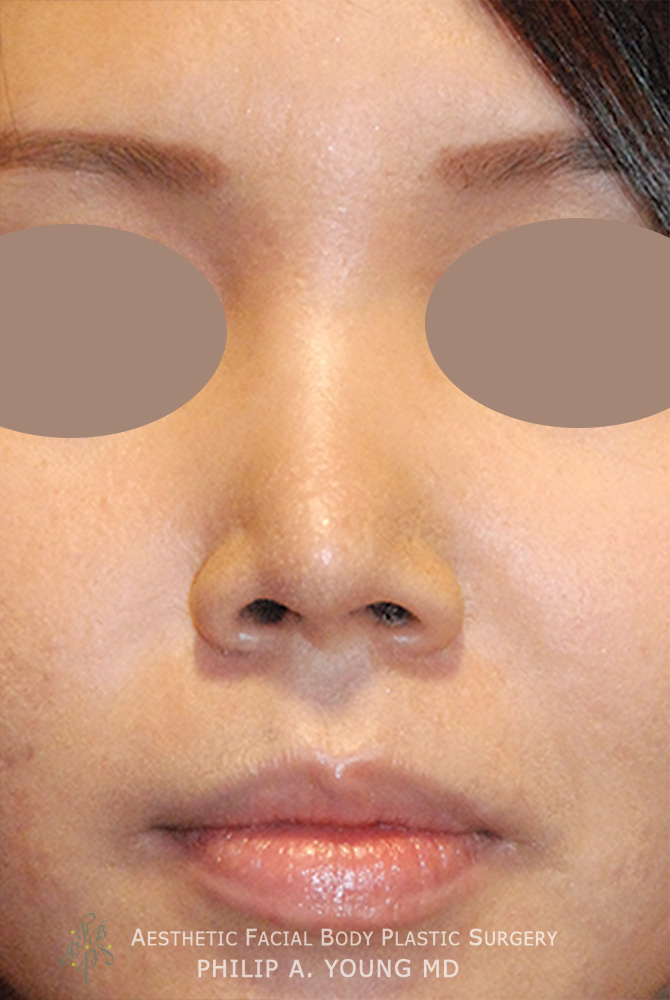
Nasal Alae: This is the soft tissue portion of the nose that covers the nostril.
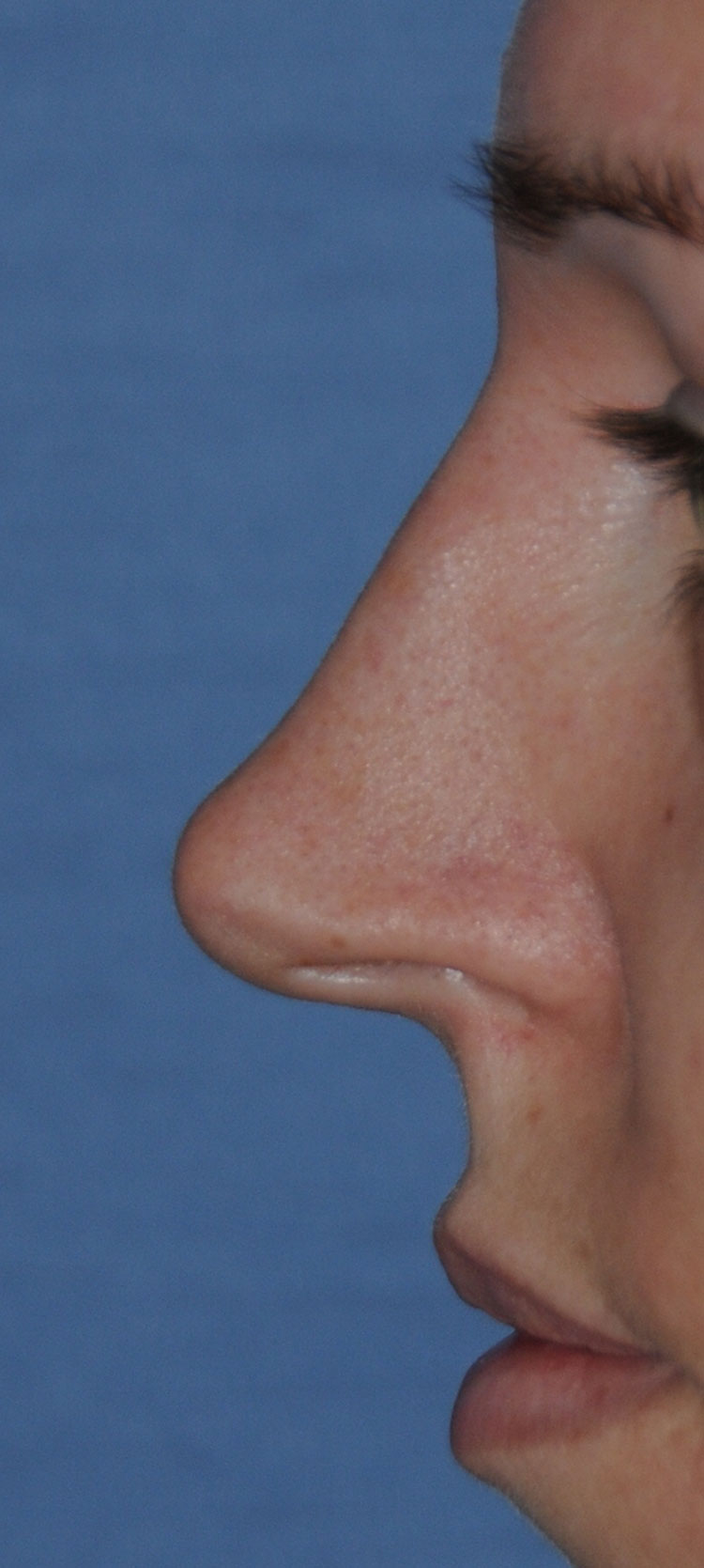
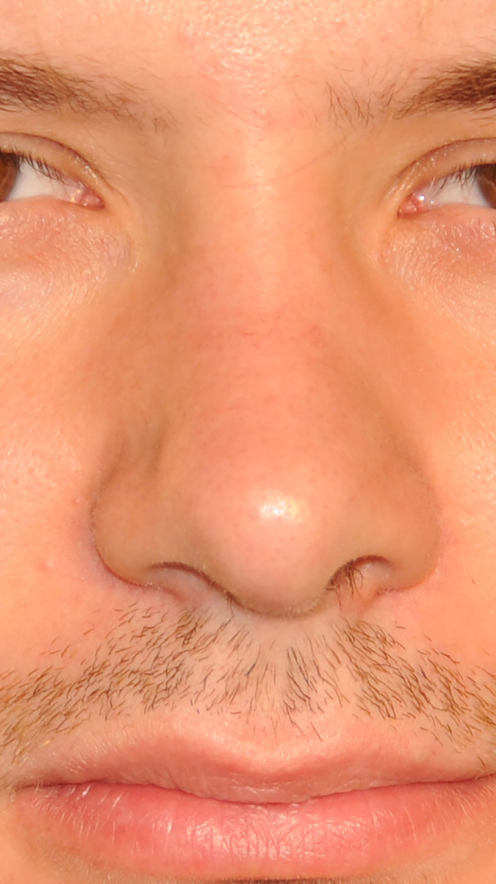
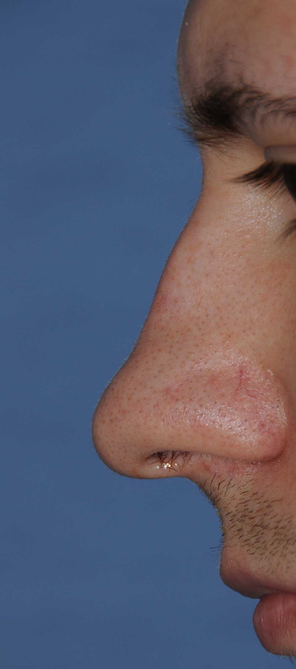
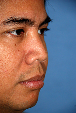
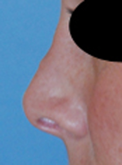
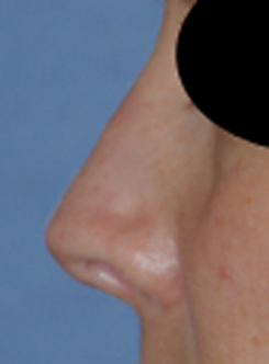
Nasal Bridge: is the highlight of the nose from the Radix | Nasion to the Nasal Tip. It is the plane of this portion of the nose that reflects the most light. The nasal sidewall is next to the nasal bridge and is shadowed more and the increased shadowing distinguishes the nasal sidewall from the nasal bridge.
Nasal Tip: is the most prominent part of the nose at the lowest part of the nose. In general, it is the part of the nose that stands out the most and is the center point of the whole nose section and plays a major role in facial beauty because it determines the center of the nose.
Radix: Is the lowest part of the nose in between the eyes and is the bony landmark. Whereas the Nasion is the soft tissue lowest point of the Nose and the surface representation of the Radix.
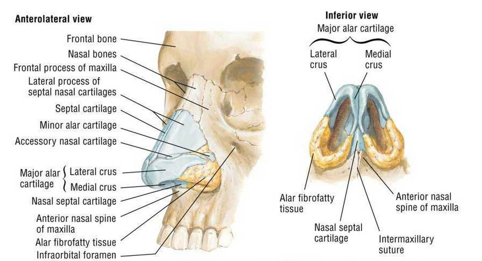
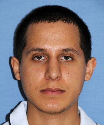
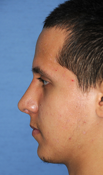
Frontal Bone: Covers the whole forehead and the frontal lobe of the brain. The glabella is the part of the frontal bone that is above and central from the eyes. The frontal bone attaches to the nasal bones
Nasal Bone: The nasal bones comprise of the upper third of the nose. The nose is broken up into thirds. The upper third is thought of as made up of bone. The middle third is made up of the upper lateral cartilages. The lower third is made up of the lower lateral cartilages
Frontal Process of the Maxilla: Is part of the maxillary bone that is attached to the nasal bones and the frontal bone. This along with the nasal bones are often involved in osteotomies that describe the cutting through bone that helps manipulate the upper third to either narrow this section of the nose or widen it.
Septal Cartilage: Is the cartilage structure that reaches the nasal bridge and is mainly the central part of the bridge that also extends far into the nasal cavity. The septal cartilage is essential to the strength of the nose and determines the projection of the nose and much of the shape of the nose. A crooked nose is highly dependent on how you treat the septal cartilage.
Upper Lateral Cartilages: Is named above as the lateral process of the septal cartilage. In the rhinoplasty world, this is commonly referred to as the upper lateral cartilages. How you change this area will determine how the middle third or middle part of the nose will look. The relation of the upper lateral cartilages and the septum can play a big factor in your nasal airway.
Lower Lateral Cartilages: Is named above the Major Alar Cartilages. In the Rhinoplasty world they are mainly referred to as the lower lateral cartilages. It is further broken up into the lateral crus | segment, middle or intermediate crus | segment, and medial crus | segment. The lateral crus and how it is position will determine a big part in how the tip will look. If it is position high closer to the septum in a cephalic orientation, or more towards the top of the head, the tip will be bigger and the nostrils will collapse easier and usually the nostril rim will be more upward curled or snarled. Also the thickness of the lower lateral cartilages will determine how big the tip is. Most refinement of the tip cartilages involve narrowing the lateral crus, specifically the top part.
Alae: Is named the Alar Fibro Fatty tissue above. The Alae is the soft tissue that covers the nostril and is made up of soft fatty tissue and no cartilage. Changing this part of the nose can narrow the bottom of the nose for nostrils that are flaring and wide.
Below is an intraoperative view of the nose:
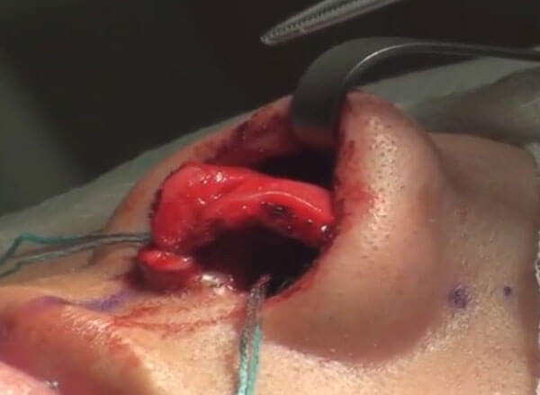
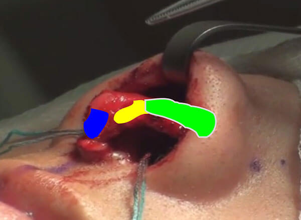
The lower lateral cartilages are shown with and without the color coding. The area outline in blue shows the position of the medial crura. The yellow the intermediate crura. The green the Lateral crura of the lower lateral cartilages. The medial crura and both lateral crura make up the tripod of the nasal tip. This is a very traditional concept of the nasal tip and how lengthening or shortening either tripod will change the position of the tip.
Below are pictures of the inside of the nose:
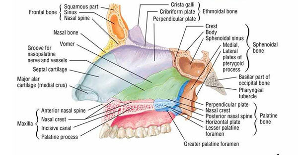
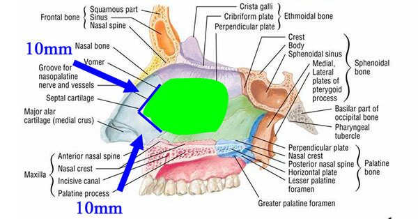
Ethmoid Bone: Is the bony part of the septum that is superior in location. It forms a major part of the medial portion of the orbital cavity as well as the septum seen here. In Rhinoplasty, often times the septum is harvested leaving behind dorsal and caudal struts of 10mm as shown in the bottom picture for nasal support. The ethmoid along with the septum is part of this harvest. This tissue can serve to help with the rhinoplasty for structural support.
Vomer Bone: is the bone that attaches to a portion of the maxilla and the palatine bone that forms the inferior bony part of the septum. It attaches to the ethmoid bone superiorly as shown. The vomer bone is also used for grafting material.
Maxilla: the maxilla forms a major part of the cheek as well as the anterior part of the palate . As seen before we saw that it contributed to the part of the nasal bones in the sense that it was the lateral part of the bony portion of the upper third, while most of it was made up of the nasal bones. The maxilla makes part of the lower portion of the septum anteriorly as shown above.
Palatine bone: this bone makes up the bottom of the septum more posteriorly attaching to the maxillary septum and maxillary palatine process and the vomer.
Orientation Anatomical Terms: Below are some words that help define how Rhinoplasty Surgeons refer to anatomical elements:
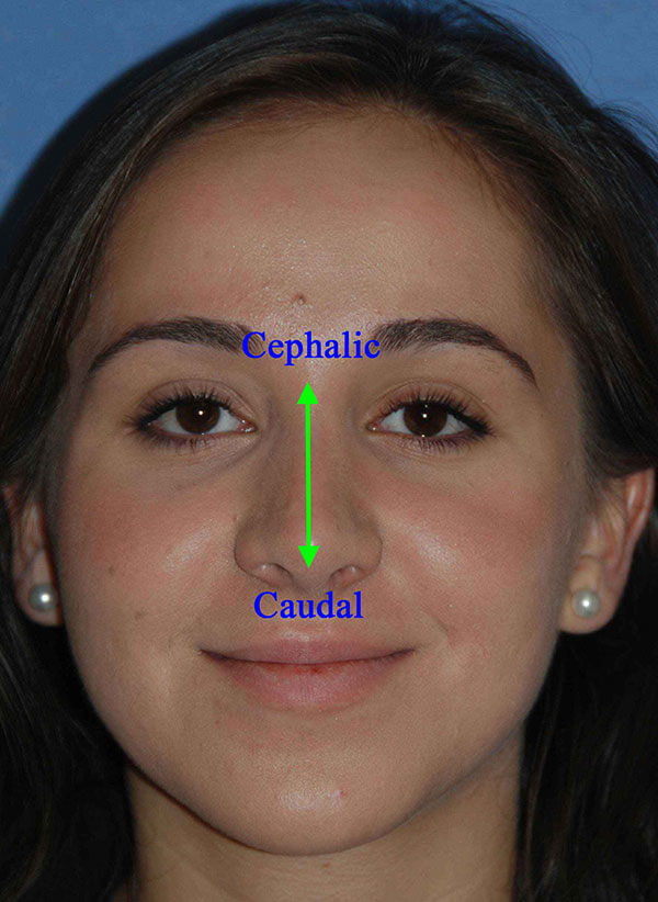
Cephalic: Means something related to the head. So with the lower lateral cartilage, we often remove the cephalicportion to refine the tip. Often, the cephalic portion creates the bulbousity of the tip or the roundness of the tip. The blue part outlined shows the cephalic portion of the lower lateral cartilages:
Caudal: In contrast, the caudal end is toward the feet. When you remove the caudal portion of the septum’s caudal end you can shorten the nose. Depending on how you shorten the caudal septum you can rotate or derotate the tip. But when you shorten the caudal septum you will always shorten the nose in some way. The picture below shows the caudal and dorsal struts of the septum. Usually you want to keep more than 10mm of the dorsal and caudal struts.
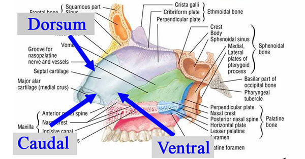
Dorsum or Dorsal: refers to the back of any anatomical or structural thing. The dorsum is a noun and dorsal is the adjective. We often preserve more than 10mm of dorsal strut to support the nose or there could be a saddle nose deformity where the dorsum collapses ventrally. This is the same for the caudal strut where more than 10mm is preserved for strength.
Ventral: refers to the belly of the anatomical structure.
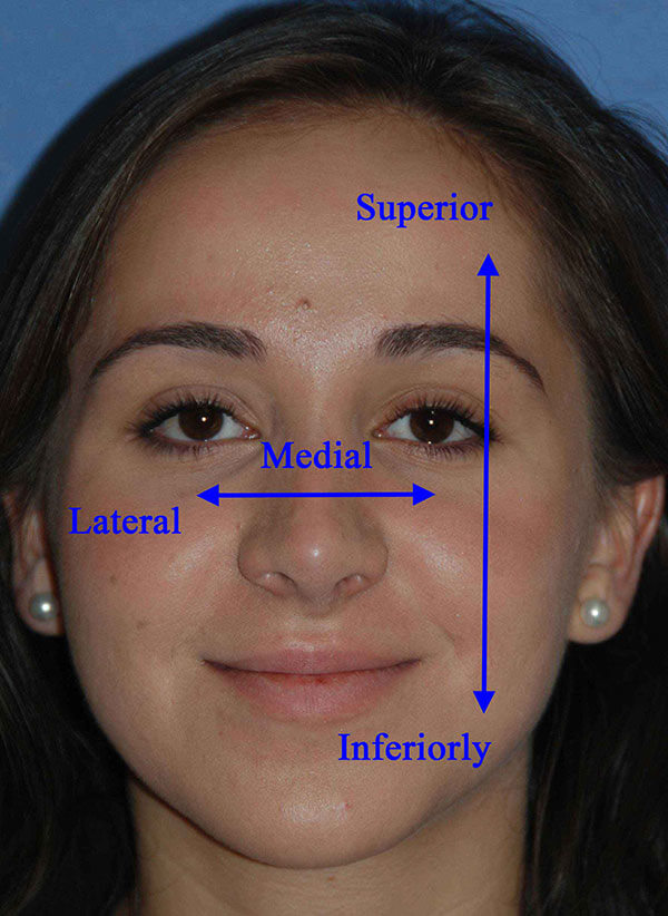
Superior: refers to things skyward
Inferior: refers to things closer to the earth’s core
Medial: refers to anything toward midline
Lateral: refers to anything away from the midline


 Patient Reviews
 Cards From Patients
 Share Your Story